|
|
| Table of Contents |

| Abbreviated Injury Score (AIS) for Inhalation Injury | RADS (Radiologist’s Score) for Smoke Inhalation Injury |
%20for%20Inhalation%20Injury.jpg) |
%20for%20Smoke%20Inhalation%20Injury.jpg) |
| Access Calculator | Access Calculator |
| Read More | Read More |
Why and When to Use, and Next Steps
|
Why to Use
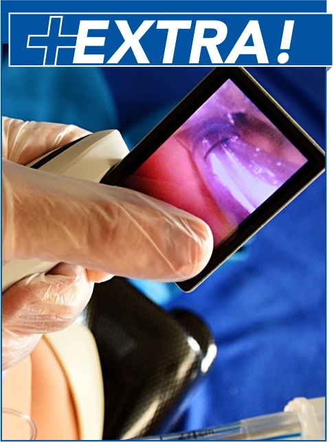
When to Use Use the AIS for adult patients with suspected inhalation injury who are undergoing flexible bronchoscopy. Next Steps
|
Pujan H. Patel, MD
Division of Pulmonary, Critical Care, and Sleep Medicine
Saint Louis University Hospital
St. Louis, Missouri
High AIS severity alone should not dictate management decisions, which should be made in conjunction with a patient’s history, physical examination, and laboratory findings.
Macroscopic manifestations of airway abnormalities may be delayed, falsely reassuring the clinician that inhalation injury has not occurred (Hunt 1975).
The AIS criteria were first proposed by Endorf and Gamelli in 2007. The purpose of their study was to identify whether the severity of inhalation injury correlated better with pulmonary parameters (eg, lung compliance, PaO2:FiO2 ratio) and acute fluid resuscitation requirement than with bronchoscopically assessed inhalation injury severity. They retrospectively reviewed 80 adult patients with suspected inhalation injury who required intubation, mechanical ventilation, and flexible bronchoscopy within the first 24 hours of admission. AIS criteria were used to separate the patients into 2 groups of bronchoscopic inhalation injury: a group with grades of 0 and 1, and a group with grades of 2, 3, and 4. Characteristics such as fluid resuscitation requirements, initial oxygenation, lung compliance, and duration of mechanical ventilation were compared between the 2 groups; however, only decreased survival correlated with bronchoscopic severity (P = .03).
Hassan et al (2010) also found a significant increase (P < .01) in mortality with bronchoscopic severity. They did not use the AIS criteria.
Since 2007, several studies have used the AIS to try to tease out a clear relationship between the bronchoscopic grade of injury and a range of clinical outcomes, with varied results. For instance, in contrast to Endorf and Gamelli’s 2007 study, studies by Albright et al (2012), Mosier et al (2012), and Spano et al (2016) found that an increasing AIS grade did not have a significant effect on mortality, with P values of 0.21, 0.10, and 0.15, respectively.
Albright et al did show that increasing severity was associated with longer ventilator days (P = .036) and ICU stays (P = .04). Mosier et al (2012) noted a significant association between AIS grade severity and the development of ARDS at 24 hours (P < .01).
Frederick W. Endorf, MD and Richard L. Gamelli, MD
Read more about Dr. Endorf and Dr.Gamelli.
Original/Primary Reference
Validation
Other References
Copyright © MDCalc • Reprinted with permission.
Why and When to Use, and Next Steps
|
Why to Use Currently, no single tool accurately and reliably risk stratifies and prognosticates outcomes for patients with smoke inhalation injury. The RADS can be a useful adjunct to determine the severity of inhalational injury to the lungs. A multicenter prospective cohort study sponsored by the American Burn Association is currently underway, with the goal of developing a scoring system for inhalation injury based on clinical, radiographic, bronchoscopic, and biochemical parameters. When to Use
Next Steps
|
Pujan H. Patel, MD
Division of Pulmonary, Critical Care, and Sleep Medicine
Saint Louis University Hospital
St. Louis, Missouri
The RADS should be used as an adjunct to clinical history, examination, bronchoscopy, and arterial blood gas data to determine the full clinical picture.
As always, clinical judgment is paramount. Management decisions should not be based solely on the RADS.
The RADS tool was developed from an ovine study of 20 anesthetized sheep who were intubated, exposed to wood smoke, and then underwent CT scans of the thorax at 6, 12, and 24 hours after exposure (Park 2003). The study raised several questions, including whether smoke inhalation from the combustion of materials other than wood would behave in the same way; whether a normal CT result would be sufficient to rule out significant injury; and how the score would perform in direct comparison to better-established diagnostic tools such as fiberoptic bronchoscopy.
Oh et al conducted a retrospective study of 43 patients (25 with inhalation injury and 19 without); using multiple logistic regression analysis, they found that inhalation injury on bronchoscopy correlated with an 8.3-fold increase in a composite endpoint of pneumonia, acute lung injury/acute respiratory distress syndrome, and death. Positive bronchoscopy in conjunction with a RADS > 8 was correlated with a 12.7-fold increase in the composite endpoints.
We are not aware of any studies looking at interrater reliability of the scoring system.
A prospective clinical trial is currently underway to help answer many of the questions that have been raised. Preliminary clinical data from the Inhalation Severity Injury Scoring System trial demonstrated a positive correlation between the RADS and ventilator days.
John S. Oh, MD
Original/Primary Reference
Other References
Copyright © MDCalc • Reprinted with permission.
Karalynn Otterness, MD; Christine Ahn, MD
Alex Manini, MD, MS, FACMT, FAACT; Lewis S. Nelson, MD
March 1, 2018
Emergency Medicine Practice • CONTINUE READING
Access every issue, our complete clinical pathway library, and earn up to 190 CME credits with an annual subscription.
Stay current with a new Emergency Medicine Practice issue every month, plus unlimited access to our complete issue library, all Interactive Clinical Pathways, and up to 190 CME credits.
Accredited By


Our Partners


 678-366-7933