Table of Contents
Rash and fever are some of the most common chief complaints presenting in emergency medicine. The evaluation of skin rashes in the febrile pediatric patient includes a broad differential diagnosis and utilizing the signs and symptoms to identify red flags, such as hemodynamic instability, erythroderma, desquamation, petechiae/purpura, mucous membrane involvement, and severe pain, in the history and physical examination that require a high index of suspicion for worrisome disease. This review addresses characteristics of common rashes, such as roseola and scarlet fever, and more rare, potentially life-threatening rashes, such as meningococcemia and toxic shock syndrome, that can be used to guide management and treatment, and improve patient outcomes. You will learn:
Characteristics that distinguish benign versus life-threatening causes of skin rash in febrile pediatric patients
Examination findings that will identify red flags in the history and physical examination
To include less commonly seen, but dangerous, conditions, and those that could be potential public health threats in the differential diagnosis of skin rash in febrile patients
To utilize the history and physical examination findings to correctly identify patients who can be discharged, or those who require diagnostic testing for further evaluation
Appropriate treatment, disposition, and follow-up recommendations for the pediatric patient with a skin rash and fever
-
Abstract
-
Case Presentations
-
Introduction
-
Critical Appraisal of the Literature
-
Etiology and Pathology
-
Differential Diagnosis
-
Prehospital Care
-
Emergency Department Evaluation
-
History
-
Physical Examination
-
Skin Examination
-
Additional Body Systems Examination
-
Diagnostic Studies
-
Management of Specific Etiologies of Rash and Fever
-
Maculopapular Rashes
-
Measles
-
Erythema Infectiosum (Fifth Disease)
-
Roseola
-
Acute Rheumatic Fever
-
Vesicular Rashes
-
Varicella
-
Hand, Foot, and Mouth Disease
-
Erythematous Rashes
-
Kawasaki Disease
-
Scarlet Fever
-
Toxic Shock Syndrome
-
Staphylococcal Scalded Skin Syndrome
-
Petechial/Purpuric Rashes
-
Meningococcal Disease
-
Henoch-Schönlein Purpura
-
Special Populations
-
Pregnant Patients
-
Unvaccinated Patients
-
Immunocompromised Patients
-
Neonates
-
Patients With Predisposition to Hemolytic Anemia
-
Controversies and Cutting Edge
-
Blood Cultures
-
Corticosteroid Use in Patients With Henoch-Schönlein Purpura and Other Rashes
-
Disposition
-
Summary
-
Time- and Cost-Effective Strategies
-
Risk Management Pitfalls for Pediatric Patients With Rash and Fever
-
Case Conclusions
-
Clinical Pathway for Emergency Department Management of Rash and Fever in the Pediatric Patient
-
Tables and Figures
-
Table 1. Common, Non–Life-Threatening Diagnoses and “Can’t Miss,” Life-Threatening Diagnoses
-
Table 2. Common Exanthem Characteristics Seen in Pediatric Patients
-
Table 3. Differential Diagnosis Based on Rash Morphology
-
Table 4. Rash Morphologies
-
Table 5. Nondermatological Physical Examination Findings by Disease/Condition
-
Table 6. Recommended Diagnostic Studies Based on Suspected Etiology
-
Table 7. Modified Jones Criteria for Diagnosing Acute Rheumatic Fever
-
Table 8. Differentiating Staphylococcal and Streptococcal Toxic Shock Syndrome
-
Table 9. TORCH Diseases
-
Figure 1. Morbilliform Rash of Measles
-
Figure 2. Rash of Erythema Infectiosum (Fifth Disease)
-
Figure 3. Viral Exanthema of Roseola
-
Figure 4. Vesicular, Multistage Rash of Varicella
-
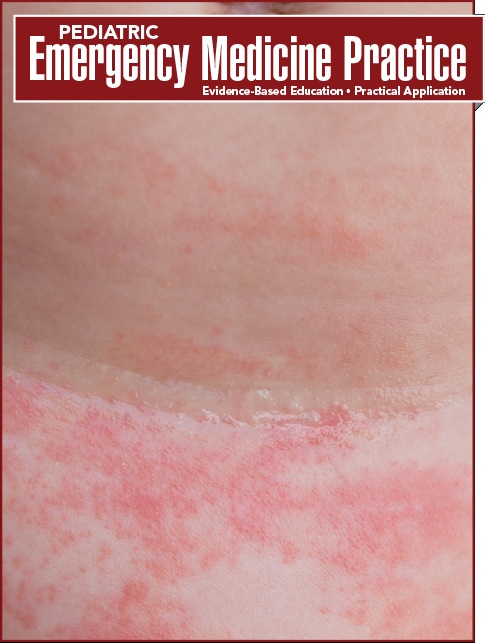
Figure 5. Rash of Scarlet Fever
-
Figure 6. Sloughing Skin of Staphylococcal Scalded Skin Syndrome
-
Figure 7. Meningococcemia Lesions
-
References
Abstract
Rash and fever are some of the most common chief complaints presenting to the emergency department. The evaluation of rashes in the febrile pediatric patient includes a broad differential diagnosis and use of the history and physical examination to identify red flags, such as hemodynamic instability, erythroderma, desquamation, petechiae/purpura, mucous membrane involvement, and severe pain, that should increase suspicion for worrisome disease. This issue reviews characteristics of common rashes as well as rarer, potentially life-threatening rashes, to guide management and treatment and improve patient outcomes.
Case Presentations
You arrive to a busy afternoon shift in the ED. Your first patient is a 1-year-old boy with rhinorrhea, congestion, cough, and 3 days of fever up to 39.4°C (103°F), measured rectally. His parents state that he has been playful at home and continues to eat and drink normally. They have been giving him acetaminophen and ibuprofen sporadically, but today he developed a generalized rash, and they became concerned. His vital signs are as follows: temperature, 38.7°C (101.7°F); heart rate, 135 beats/min; and blood pressure, 85/55 mm Hg. On examination, the rash is macular, erythematous, and blanching, but his eyes and mouth appear normal.
In the next room, there is a 3-year-old boy with a similar history who had mild rhinorrhea and a low-grade fever of 38.1°C (100.5°F) at home. His parents are concerned that he has been complaining of pain in his legs, on which they have noticed dark spots. He has continued to drink well, though he has been eating slightly less. His vitals signs are as follows: temperature, 37.5°C (99.5°F); heart rate, 120 beats/min; and blood pressure, 90/60 mm Hg. You observe some nonblanching spots on his lower extremities and buttocks, as well as mild edema and tenderness of his knees and ankle, but the boy is still able to bear weight with a mild limp.
Before you finish examining the boy, a nurse asks you to see another patient who she says does not look well. The patients is a 9-year-old girl with a history of ulcerative colitis who was sent from her pediatrician's office. She has had 4 days of sore throat and low-grade fever at home, and her parents assumed she had a cold. She tested positive for strep throat at her appointment today. Her vital signs are as follows: temperature, 38.5°C (101.3°F); heart rate, 126 beats/min; and blood pressure, 85/60 mm Hg. On examination, her skin appears diffusely erythematous as if she has a severe sunburn. These 3 patients all presented with dermatologic findings and fever. How do you determine which patients are truly ill, and which are not? Are there any red flags for recognizing rashes that could be life-threatening? Are there any key components to the history that are concerning? Do all of the patients need laboratory workup, or can you safely offer supportive care? Should any of these children be on isolation, either for their safety or for the safety of others?
Introduction
According to a 2015 United States Centers for Disease Control and Prevention (CDC) report, the single most common chief complaint for children aged < 15 years was fever, and the fifth most common was skin rash.1 When paired, fever and rash may create a diagnostic dilemma for the emergency clinician. Although many relatively benign conditions present with these symptoms, some life-threatening disease states will also present as a rash in a febrile patient. Since the differential diagnosis is broad, most management decisions will be directed by key components of the history and physical examination, and any red flags. These findings should prompt consideration of diseases that would be severely detrimental to the child’s health if missed. Some rashes, such as varicella, measles, and rubella, may represent a public health concern. Diseases such as these have become slightly more prevalent in the United States, due to caregiver concerns regarding vaccinations. Rates of meningococcal disease have decreased; however, this disease does have high rates of morbidity and mortality.2
This issue of Pediatric Emergency Medicine Practice reviews various disease states, from benign to life-threatening, that can present as a fever with rash in a child. Workup, treatment, and disposition recommendations are provided based on key features of the history and physical examination.
Critical Appraisal of the Literature
A literature search was performed using PubMed. Search terms included fever, rash, viral exanthema, measles, scarlet fever, rubella, varicella, roseola, parvovirus, lyme disease, erythema migrans, Rocky Mountain spotted fever, acute rheumatic fever, erythema marginatum, Kawasaki disease, Henoch-Schönlein purpura, HSP and steroids, erythroderma, staphylococcal scalded skin syndrome, meningococcal disease, Neisseria meningitidis, purpura and fever, and toxic shock syndrome.
Multiple reviews and case reports were found, but, overall, evidence-based literature and original research was scarce. Information from the World Health Organization (WHO) and the CDC was also incorporated, as well as information from textbooks in infectious disease, emergency medicine, and pediatrics specialties.
Risk Management Pitfalls for Pediatric Patients With Rash and Fever
1. “This child had a sore throat with exudate and a rash. I thought it must be scarlet fever and gave him antibiotics.”
Prior to prescribing antibiotics, the characteristics of the rash, associated symptoms, history, and physical examination findings should be taken into account to develop the differential diagnosis. Many viruses can cause skin rash and exudative pharyngitis that would not benefit from antibiotics (eg, mononucleosis).
2. “The patient did not have strep throat recently, so it can’t be acute rheumatic fever.”
Often, children have an episode of pharyngitis and do not have a rapid strep test performed to make the diagnosis of “strep throat.” Additionally, the symptoms of ARF appear after a 2- to 3-week latent period following the streptococcal pharyngitis, so prior infection may not be evident at the time of presentation.
3. “The patient has had 5 days of fever and now has a rash; this is probably just a viral exanthem.”
In the setting of 5 days of fever, Kawasaki disease should be considered, especially in infants who may not have the classic presentation of fever ≥ 5 days and at least 4 out of the 5 following clinical features: (1) conjunctival injection, (2) mucous membrane changes (bright red, cracked lips, strawberry tongue), (3) changes in peripheral extremities (usually a later finding), (4) polymorphous rash, and (5) cervical lymphadenopathy.
Tables and Figures
References
Evidence-based medicine requires a critical appraisal of the literature based upon study methodology and number of subjects. Not all references are equally robust. The findings of a large, prospective, randomized, and blinded trial should carry more weight than a case report.
To help the reader judge the strength of each reference, pertinent information about the study is included in bold type following the reference, where available. In addition, the most informative references cited in this paper, as determined by the author, are highlighted.
-
Rui P, Kang K. National hospital ambulatory medical care survey: 2015 emergency department summary tables. Accessed December 15, 2019. (CDC data)
-
Adams D, Thomas K, Jajosky R, et al. Summary of notifiable infectious diseases and conditions - United States, 2014. MMWR Morb Mortal Wkly Rep. 2016;63(54):1-152. (CDC data)
-
Aber C, Alvarez Connelly E, Schachner LA. Fever and rash in a child: when to worry? Pediatr Ann. 2007;36(1):30-38. (Review)
-
United States Centers for Disease Control and Prevention. Measles. Accessed December 15, 2019. (CDC data)
-
Moss WJ. Measles. Lancet. 2017;390(10111):2490-2502. (Review)
-
Dyer JA. Childhood viral exanthems. Pediatr Ann. 2007;36(1):21-29. (Review)
-
World Health Organization. Measles vaccines: WHO position paper, April 2017. Weekly Epidemiological Record. 2017;92(17):205-228. (Position paper, review)
-
Buchanan R, Bonthius DJ. Measles virus and associated central nervous system sequelae. Semin Pediatr Neurol. 2012;19(3):107-114. (Review)
-
O’Grady JS. Fifth and sixth diseases: more than a fever and a rash. J Fam Pract. 2014;63(10):E1-E5. (Review)
-
Smith PT, Landry ML, Carey H, et al. Papular-purpuric gloves and socks syndrome associated with acute parvovirus B19 infection: case report and review. Clin Infect Dis. 1998;27(1):164-168. (Case report, review)
-
Watkins J. Red faces: making the diagnosis. Pract Nursing. 2008;19(1):31-34. (Review)
-
Mullins TB, Krishnamurthy K. Roseola infantum (exanthema subitum, sixth disease). StatPearls. StatPearls Publishing LLC; Treasure Island, FL: 2018. (Review)
-
Khan A, Sutcliffe N, Jawad AS. An old disease re-emerging: acute rheumatic fever. Clin Med (Lond). 2018;18(5):400-402. (Case report)
-
Rhodes KL, Rasa MM, Yamamoto LG. Acute rheumatic fever: revised diagnostic criteria. Pediatr Emerg Care. 2018;34(6):436-440. (Review)
-
Gewitz MH, Baltimore RS, Tani LY, et al. Revision of the Jones Criteria for the diagnosis of acute rheumatic fever in the era of Doppler echocardiography: a scientific statement from the American Heart Association. Circulation. 2015;131(20):1806-1818. (Practice guideline)
-
Gerber MA, Baltimore RS, Eaton CB, et al. Prevention of rheumatic fever and diagnosis and treatment of acute streptococcal pharyngitis: a scientific statement from the American Heart Association Rheumatic Fever, Endocarditis, and Kawasaki Disease Committee of the Council on Cardiovascular Disease in the Young, the Interdisciplinary Council on Functional Genomics and Translational Biology, and the Interdisciplinary Council on Quality of Care and Outcomes Research: endorsed by the American Academy of Pediatrics. Circulation. 2009;119(11):1541-1551. (Practice guideline)
-
Sika-Paotonu D, Beaton A, Raghu A, et al. Acute rheumatic fever and rheumatic heart disease. In: Ferretti JJ, Stevens DL, Fischetti VA, eds. Streptococcus pyogenes: Basic Biology to Clinical Manifestations. Oklahoma City, OK: University of Oklahoma Health Sciences Center; 2016. (Textbook chapter)
-
Global Burden of Disease Collaborative Network. Global Burden of Disease Study 2017 (GBD 20176) Data Resources. Institute for Health Metrics and Evaluation (IHME): Seattle, WA. Accessed December 15, 2019. (Population study)
-
Heininger U, Seward JF. Varicella. Lancet. 2006;368(9544): 1365-1376. (Review)
-
National Center for Immunization and Respiratory Diseases. Chickenpox (varicella). United States Centers for Disease Control and Prevention; 2018. Accessed December 15, 2019. (CDC data)
-
Kim KS, Hufnagel G, Chapman NM, et al. The group B coxsackieviruses and myocarditis. Rev Med Virol. 2001;11(6):355-368. (Review)
-
Xing W, Liao Q, Viboud C, et al. Hand, foot, and mouth disease in China, 2008-12: an epidemiological study. Lancet Infect Dis. 2014;14(4):308-318. (Retrospective, population-based study; 267,942 confirmed cases)
-
Dietz SM, van Stijn D, Burgner D, et al. Dissecting Kawasaki disease: a state-of-the-art review. Eur J Pediatr. 2017;176(8):995-1009. (Review)
-
Kato H, Ichinose E, Yoshioka F, et al. Fate of coronary aneurysms in Kawasaki disease: serial coronary angiography and long-term follow-up study. Am J Cardiol. 1982;49(7):1758-1766. (Follow-up study; 42 patients)
-
Holman RC, Belay ED, Christensen KY, et al. Hospitalizations for Kawasaki syndrome among children in the United States, 1997-2007. Pediatr Infect Dis J. 2010;29(6):483-488. (Retrospective analysis)
-
McCrindle BW, Rowley AH, Newburger JW, et al. Diagnosis, treatment, and long-term management of Kawasaki disease: a scientific statement for health professionals from the American Heart Association. Circulation. 2017;135(17):e927-e999. (Practice guideline)
-
Allmon A, Deane K, Martin KL. Common skin rashes in children. Am Fam Physician. 2015;92(3):211-216. (Review)
-
Brinker A. Scarlet fever. N Engl J Med. 2017;376(20):1972. (Review)
-
Wessels MR. Pharyngitis and scarlet fever. In: Ferretti JJ, Stevens DL, Fischetti VA, eds. Streptococcus pyogenes: Basic Biology to Clinical Manifestations. Oklahoma City, OK: University of Oklahoma Health Sciences Center; 2016. (Textbook chapter)
-
Parker MW, Shah SS. Infectious diseases. In: Shah BR, Mahajan P, Amodio J, et al, eds. Atlas of Pediatric Emergency Medicine. 3rd ed. New York, NY: McGraw-Hill Education. (Textbook chapter)
-
van Driel ML, De Sutter AI, Keber N, et al. Different antibiotic treatments for group A streptococcal pharyngitis. Cochrane Database Syst Rev. 2013;(4):CD004406. (Systematic review and meta-analysis; 19 trials, 5839 participants)
-
Gottlieb M, Long B, Koyfman A. The evaluation and management of toxic shock syndrome in the emergency department: a review of the literature. J Emerg Med. 2018;54(6):807-814. (Review)
-
Peterson ML. A long road to a preventative for toxic shock syndrome. Lancet Infect Dis. 2016;16(9):985-986. (Commentary)
-
Lappin E, Ferguson AJ. Gram-positive toxic shock syndromes. Lancet Infect Dis. 2009;9(5):281-290. (Review)
-
United States Centers for Disease Control and Prevention. Toxic shock syndrome (other than streptococcal) (TSS). 2011 Case Definition. Accessed December 15, 2019. (CDC data)
-
United States Centers for Disease Control and Prevention. Streptococcal toxic shock syndrome (STSS) (Streptococcus pyogenes). 2010 Case Definition. Accessed December 15, 2019.(CDC data)
-
Grama A, Marginean OC, Melit LE, et al. Staphylococcal scalded skin syndrome in child. A case report and a review from literature. J Crit Care Med (Targu Mures). 2016;2(4):192-197. (Case report, review)
-
Bukowski M, Wladyka B, Dubin G. Exfoliative toxins of Staphylococcus aureus. Toxins (Basel). 2010;2(5):1148-1165. (Review)
-
Mishra AK, Yadav P, Mishra A. A systemic review on staphylococcal scalded skin syndrome (SSSS): a rare and critical disease of neonates. Open Microbiol J. 2016;10:150-159. (Review)
-
Thomas AE, Baird SF, Anderson J. Purpuric and petechial rashes in adults and children: initial assessment. BMJ. 2016;352:i1285. (Review)
-
United States Centers for Disease Control and Prevention. Epidemiology and Prevention of Vaccine-Preventable Diseases. Hamborsky J, Kroger A, Wolfe S, eds. 13th ed. Washington D.C.: Public Health Foundation; 2015. (Textbook)
-
Olbrich KJ, Muller D, Schumacher S, et al. Systematic review of invasive meningococcal disease: sequelae and quality of life impact on patients and their caregivers. Infect Dis Ther. 2018;7(4):421-438. (Systematic review; 31 studies)
-
Waterfield T, Lyttle MD, Fairley D, et al. The “Petechiae in children” (PiC) study: evaluating potential clinical decision rules for the management of feverish children with non-blanching rashes, including the role of point of care testing for procalcitonin & Neisseria meningitidis DNA - a study protocol. BMC Pediatr. 2018;18(1):246. (Prospective diagnostic accuracy study)
-
Ang E, Sundel R. Henoch-Schönlein purpura. In: Chang V, Zaoutis L, eds. Comprehensive Pediatric Hospital Medicine. 2nd ed. New York, NY: McGraw-Hill Education; 2017:832-835. (Textbook chapter)
-
Friedman M, Mendoza C. Petechiae and purpura. In: Tenenbein M, Macias C, Sharieff G, et al, eds. Strange and Schafermeyer’s Pediatric Emergency Medicine. 5th ed. New York, NY: McGraw-Hill Education; 2018:549-552. (Textbook chapter)
-
McCarthy HJ, Tizard EJ. Clinical practice: diagnosis and management of Henoch-Schönlein purpura. Eur J Pediatr. 2010;169(6):643-650. (Review)
-
Saulsbury FT. Henoch-Schönlein purpura in children. Report of 100 patients and review of the literature. Medicine (Baltimore). 1999;78(6):395-409. (Case reports, review; 100 patients)
-
Burks M, Usatine R. Henoch-Schönlein purpura. In: Usatine R, Sabella C, Smith M, et al., eds. The Color Atlas of Pediatrics. 1st ed. New York, NY: McGraw-Hill Pediatrics; 2015:1012-1017. (Textbook chapter)
-
Rajput R, Sehgal A, Jain D, et al. Acute parvovirus B19 infection leading to severe aplastic anemia in a previously healthy adult female. Indian J Hematol Blood Transfusion. 2012;28(2):123-126. (Case report)
-
Woods-Hill CZ, Fackler J, Nelson McMillan K, et al. Association of a clinical practice guideline with blood culture use in critically ill children. JAMA Pediatr. 2017;171(2):157-164. (Retrospective cohort study; 2356 patients)
-
Ronkainen J, Koskimies O, Ala-Houhala M, et al. Early prednisone therapy in Henoch-Schönlein purpura: a randomized, double-blind, placebo-controlled trial. J Pediatr. 2006;149(2):241-247. (Randomized double-blind placebo-controlled trial; 171 patients)
-
Delbet JD, Hogan J, Aoun B, et al. Clinical outcomes in children with Henoch-Schönlein purpura nephritis without crescents. Pediatr Nephrol. 2017;32(7):1193-1199. (Retrospective multicenter study; 92 patients)
-
Hahn D, Hodson EM, Willis NS, et al. Interventions for preventing and treating kidney disease in Henoch-Schönlein purpura (HSP). Cochrane Database Syst Rev. 2015(8):CD005128. (Systematic review; 13 randomized controlled trials)
-
Dudley J, Smith G, Llewelyn-Edwards A, et al. Randomised, double-blind, placebo-controlled trial to determine whether steroids reduce the incidence and severity of nephropathy in Henoch-Schönlein purpura (HSP). Arch Dis Child. 2013;98(10):756-763. (Randomized controlled trial; 352 patients)
-
Jauhola O, Ronkainen J, Koskimies O, et al. Outcome of Henoch-Schönlein purpura 8 years after treatment with a placebo or prednisone at disease onset. Pediatr Nephrol. 2012;27(6):933-939. (Randomized trial; 160,138 patients)
-
Wardle AJ, Connolly GM, Seager MJ, et al. Corticosteroids for the treatment of Kawasaki disease in children. Cochrane Database Syst Rev. 2017;1:Cd011188. (Systematic review)


 678-366-7933