|
|
Jeff: Welcome back to EMplify, the podcast corollary to EB Medicine’s Emergency Medicine Practice. I’m Jeff Nusbaum, and I’m back with my co-host, Nachi Gupta. This month, after a few months of primarily medical topics, we’re talking trauma, specifically Blunt Cardiac Injury: Emergency Department Diagnosis and Management.
Nachi: With no gold standard diagnostic test and with complications ranging from simple ectopic beats to fulminant cardiac failure and death, this isn’t an episode you’ll want to miss.
Jeff: Before we begin, let me give a quick shout out to our incredible group of authors from New York -- Dr. Eric Morley, Dr. Bryan English, and Dr. David Cohen of Stony Brook Medicine and Dr. William Paolo, residency program director at SUNY Upstate. I should also mention their peer reviewers Drs. Jennifer Maccagnano and Ashley Norse of the NY institute of technology college of osteopathic medicine and UF Health Jacksonville, respectively.
Nachi: This month’s team parsed through roughly 1200 articles as well as guidelines from the eastern association for surgery in trauma also known as EAST.
Jeff: Clearly a large undertaking for a difficult topic to come up with solid evidence based recommendations.
Nachi: For sure. Let’s begin with some epidemiology, which is admittedly quite difficult without universally accepted diagnostic criteria.
Jeff: As you likely know, despite advances in motor vehicle safety, trauma remains a leading cause of death for young adults. In the US alone, each year, there are about 900,000 cases of cardiac injury secondary to trauma. Most of these occur in the setting of vehicular trauma.
Nachi: And keep in mind, that those injuries don’t occur in isolation as 70-80% of patients with blunt cardiac injury sustain other injuries. This idea of concomitant trauma will be a major theme in today’s episode.
Jeff: It certainly will. But before we get there, we have some more definitions to review - cardiac concussion and contusion, both of which were defined in a 1989 study. In this study, cardiac concussion was defined as an elevated CKMB with a normal echo, while a cardiac contusion was defined as an elevated CKMB and abnormal echo.
Nachi: Much to my surprise, though, abnormal echo and elevated ck-mb have not been shown to be predictive of adverse outcomes, but conduction abnormalities on ekgs have been predictive of development of serious dysrhythmia
Jeff: More on complications in a bit, but first, returning to the idea of concomitant injuries, in one autopsy study of nearly 1600 patients with blunt trauma - cardiac injuries were reported in 11.9% of cases and contributed to the death of 45.2% of those patients.
Nachi: Looking more broadly at the data, according to one retrospective review, blunt cardiac injury may carry a mortality of up to 44%.
Jeff: That’s scary high, though I guess not terribly surprising, given that we are discussing heart injuries due to major trauma...
Nachi: The force may be direct or indirect, involve rapid deceleration, be bidirectional, compressive, concussive, or even involve a combination of these. In general, the right ventricle is the most frequently injured area due to the proximity to the chest wall.
Jeff: Perfect, so that's enough background, let’s talk differential. As you likely expected, the differential is broad and includes cardiovascular injuries, pulmonary injuries, and other mediastinal injuries like pneumomediastinum and esophageal injuries.
Nachi: Among the most devastating injuries on the differential is cardiac wall rupture, which not surprisingly has an extremely high mortality rate. In terms of location of rupture, both ventricles are far more likely to rupture than the atria with the right atria being more likely to rupture than the left atria. Atrial ruptures are more survivable, whereas complete free wall rupture is nearly universally fatal.
Jeff: Septal injuries are also on the ddx. Septal injuries occur immediately, either from direct impact or when the heart becomes compressed between the sternum and the spine. Delayed rupture can occur secondary to an inflammatory reaction. This is more likely in patients with a prior healed or repaired septal defects.
Nachi: Valvular injuries, like septal injuries, are rare. Left sided valvular damage is more common and carries a higher mortality risk. In order, the aortic valve is more commonly injured followed by the mitral valve then tricuspid valve, and finally the pulmonic valve. Remember that valvular damage can be due to papillary muscle rupture or damage to the chordae tendineae. Consider valvular injury in any patient who appears to be in cardiogenic shock, has hypotension without obvious hemorrhage, or has pulmonary edema.
Jeff: Next on the ddx are coronary artery injuries, which include lacerations, dissections, aneurysms, thrombosis, and even MI secondary to increased sympathetic activity and platelet activity after trauma. In one review, dissection was the most commonly uncovered pathology, occurring 71% of the time, followed by thrombosis, which occured only 7% of the time. The LAD is the most commonly injured artery followed by the RCA.
Nachi: Pericardial injury, including pericarditis, effusion, tamponade, and rarely rupture, is also certainly on the differential.
Jeff: In terms of dysrhythmias, sinus tachycardia is the most common dysrhythmia, with other rhythms, including PVC / PAC / and afib being found only 1-6% of the time.
Nachi: And while conduction blocks are rare, a RBBB is the most commonly noted, followed by a 1st degree AVB.
Jeff: Though also rare, commotio cordis deserves it’s own section as its the second most common cause of death in athletes < 18 who are victims of blunt trauma. Though only studied in swine models, it’s hypothesized that the impact to the chest wall during T-wave upstroke can precipitate v-fib.
Nachi: Aortic root injuries usually occur at the insertion of the ligamentum arteriosum and isthmus. Such injuries typically result in aortic insufficiency.
Jeff: And the last pathology on the differential requiring special attention is a myocardial contusion. Again, no standard definition exists, with some diagnostic criteria including simply chest pain and increasing cardiac enzymes, and others including cardiac dysfunction, ecg abnormalities, wall motion abnormalities, and an elevation of cardiac enzymes.
Nachi: Certainly a pretty broad differential… before moving on to the work up, Jeff why don’t you get us started with prehospital care?
Jeff: Prehospital management should focus on rapid identification and stabilization of life threatening injuries with expeditious transport as longer prehospital times have been associated with increased mortality in trauma. Immediate transport to a Level I trauma center should be the highest priority for those with suspected blunt cardiac injury.
Nachi: In terms of who specifically should be transporting the patient, a Cochrane review evaluated the utility of ALS vs BLS transport in trauma. There is reasonably good data to support BLS over ALS, even when controlling for trauma severity. Moreover, when airway management is needed, advanced airway techniques by ALS crews were associated with decreased odds of survival. Regardless of who is there, the message is the same: focus not on interventions, but instead on rapid transport.
Jeff: And if it does happen to be an ALS transport crew, without delaying transport, pain management with fentanyl is both safe and reasonable and preferred over morphine. Post opiate hypotension in prehospital trauma patients is a rare but documented complication.
Nachi: And if the prehospital team is lucky enough, or maybe unlucky enough, i don’t know, to have a credentialed provider who can perform ultrasound for those suspected of having a blunt cardiac injury, the general prehospital data on ultrasound is sparse. As of now, it’s difficult to conclude if prehospital US improves care for trauma patients.
Jeff: Interestingly, the system I work in has prehospital physicians, who do carry US, but I can’t think of a major trauma where ultrasound changed any of the decisions we made.
Nachi: Right, and I think that just reinforces the main point here: there may be a role, we just don’t have the data to support it at this time.
Jeff: Great, let’s move onto ED care, beginning with the H&P.
Nachi: On history, make sure to elucidate if there is any chest pain, and if it’s onset was before or after the traumatic event. In addition, make sure to ask about dyspnea, fatigue, palpitations, and lightheadedness.
Jeff: And don’t forget to get the crash details from the EMS crew before they depart! As a side note, for anyone taking oral boards in a few months, don’t forget to ask the EMS crew for the details!!!
Nachi: A definite must for oral boards and for your clinical practice.
Jeff: In terms of the physical, tachycardia is the most common abnormality in blunt cardiac injury. In those with severe injury, you may note refractory hypotension secondary to cardiogenic shock. But don’t be reassured by normal vitals, especially in the young, who may be compensating well despite being quite ill.
Nachi: Fully undress the patient to appropriately inspect and percuss the chest wall - looking for signs of previous cardiac surgeries or pacemaker placement, as well as to auscultate for new murmurs which may be a sign of valvular injury.
Jeff: Similarly, as concomitant injuries are common, inspect the abdomen, looking for ecchymosis patterns, which often accompany blunt cardiac injury.
Nachi: Pretty standard stuff. Let’s move on to diagnostic testing.
Jeff: Lab testing should include a CBC, BMP, coags, troponin, lactate, and T&S. In one retrospective analysis, an elevated troponin and a lactate over 2.5 were predictors of mortality.
Nachi: Additionally, in patients with chest trauma, a troponin > 1.05 was associated with a greater risk for dysrhythmias and LV dysfunction.
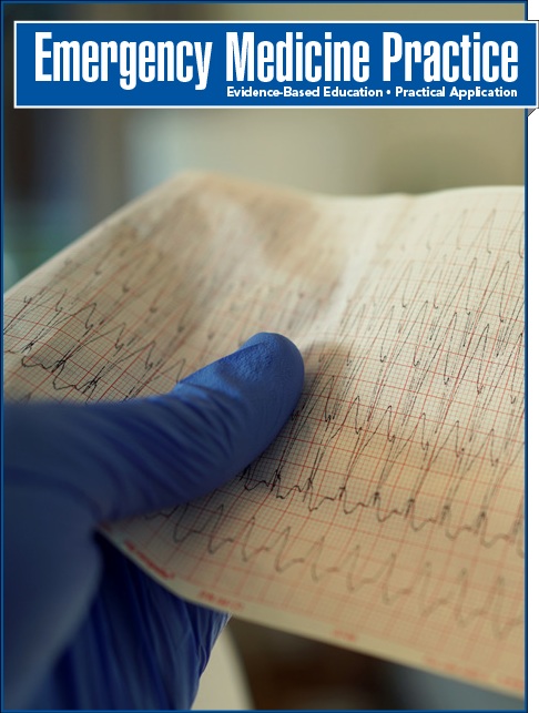
Jeff: And it likely goes without saying, but an EKG is a must on all trauma patients with suspicion for blunt cardiac injury in accordance with the EAST guidelines. New EKG findings requires admission for monitoring. Unfortunately, on the flip side, an ECG cannot be used to rule out blunt cardiac injury.
Nachi: Diving a bit deeper into the data, in a prospective study of 333 patients with blunt thoracic trauma, serial EKG and troponins at 0, 4, and 8 hours post injury had a sensitivity and specificity of 100% and 71%, respectively. However, of those with abnormal findings, all but one had them on initial testing, leading to a negative predictive value of 98%.
Jeff: Well that’s an impressive NPV and has huge implications, especially in the era of heavily monitored lengths of stay...
Nachi: Definitely. In terms of radiography, a chest x-ray should be obtained as rib fractures, hemopneumothorax, and mediastinal free air are all things you wouldn't want to miss and are also associated with blunt cardiac injury.
Jeff: Keep in mind, however, that the chest x-ray should not be seen as a test for pericardial fluid as up to 200 mL of fluid can be contained in the pericardial space and remain undetectable by chest radiograph.
Nachi: Which is why you’ll have to turn to our good friend the ultrasound, for more useful data. The data is strong that in the hands of trained Emergency Clinicians, when parasternal, apical, and subcostal views are obtained, US has an accuracy of 97.5% for pericardial effusion.
Jeff: Not only is US accurate, it’s also quick. In one RCT, the FAST exam reduced the time from arrival in the ED to operative care by 64% in the setting of trauma.
Nachi: That’s impressive -- for expediting patient care and for managing ED flow.
Jeff: Exactly. The authors do note however that hemopericardium is a rare finding, so, while not the focus of this article, the real utility of the FAST exam may be in its expanded form, the eFAST, in which a rapid bedside ultrasonographic lung exam for pneumothorax is included, as this can lead to immediate changes in management.
Nachi: And assuming you do your FAST or eFAST and have no management changing findings, CT will often be your next test.
Jeff: Yeah, EKG-gated multidetector CT can easily diagnose myocardial rupture, pneumopericardium, pericardial rupture, hemopericardium, coronary artery insult, ventricular septal defects and even valvular dysfunction. Unfortunately, CT does not perform well for the evaluation of myocardial contusions.
Nachi: This is all well and good, and certainly accurate, but let’s not forget that hemodynamically unstable trauma patients, like those with myocardial rupture, need to be in the operating room, not the CT scanner.
Jeff: An important point that should not be understated.
Nachi: And the last major testing modality to discuss is the echocardiogram.
Jeff: The echo is a fantastic test for detecting focal cardiac dysfunction often see with cardiac contusions, hemopericardium, and valve disruption.
Nachi: And it’s worth noting that transthoracic is enough, as transesophageal, despite the better images, hasn’t been shown to change management. TEE should be saved for those in whom a optimal TTE study isn’t feasible.
Jeff: Great point. And one last quick note on echo: in terms of guidelines, the EAST guidelines from 2012 specifically recommend an echo in hemodynamically unstable patients or those with a persistent new dysrhythmia without other sources of ongoing hemorrhage or neurologic etiology of instability.
Nachi: Perfect, so that wraps up testing and imaging for our blunt cardiac injury patient. Let’s move on to treatment.
Jeff: In terms of initial resuscitation, there is an ever increasing body of literature to support blood transfusion over crystalloid in patients requiring volume expansion in trauma. There are no specific guidelines for transfusion in the setting of blunt cardiac injury, so stick to your standard trauma protocols.
Nachi: It is worth noting, though, that there is literature outside of trauma for those with pericardial effusions, suggesting that those with a SBP < 100 have substantial benefit from volume expansion. So keep this in mind if your clinical suspicion is high and your trauma patient has a soft but not truly shocky blood pressure.
Jeff: Operative management, specifically ED thoracotomy is a heavily debated topic, and it’s next on our list to discuss.
Nachi: The 2015 EAST guidelines conditionally recommend ED thoracotomy for moribund patients with signs of life. The Western Trauma Association broadens the ED thoracotomy window a bit to include anyone with no signs of life but less than 10 minutes of CPR. The latter also recommend ED thoracotomy in those with refractory shock.
Jeff: Though few studies exist on the topic, in one study of 187 patients, cardiac motion on US was 100% sensitive for predicting survivors.
Nachi: Not great data, but it does support one's decision to stop any further work up should there be no cardiac activity, which is important, because the decision to pursue an ED thoracotomy is not an easy one.
Jeff: And lastly, emergent pericardiocentesis may be another option in an unstable patient when definitive operative management is not possible. But do note that pericardiocentesis is only a temporizing measure, and not definitive for cardiac tamponade.
Nachi: Treatment for dysrhythmias is standard, treat in accordance with standard ACLS protocols, as formal randomized trials on prophylaxis and treatment in the setting of blunt cardiac injury do not exist.
Jeff: Seems reasonable enough. And in the very rare setting of an MI after blunt cardiac injury, you should involve cardiology, cardiothoracic surgery, and trauma to help make important management decisions. Data is, again, lacking, but the patient likely needs percutaneous angiography for appropriate diagnosis and potentially further intervention. Definitely hold off on ASA and likely nitroglycerin, at least until significant bleeding has been ruled out.
Nachi: Yup, no style points for giving aspirin to a bleeding trauma patient. Speaking of medications, the last treatment modality to discuss here is pain control. Pain management is essential with chest injuries, as appropriate pain management has been shown to reduce mortality in pulmonary related complications.
Jeff: And in line with every acute pain consult note I’ve ever come across, a multimodal approach utilizing opioids and nonopioids is recommended.
Nachi: Perfect, so that sums up treatment, next we have one special circumstance to discuss: sternal fractures. Cardiac contusions are found in 1.8-2.4% of patients with sternal fractures, almost all of which were seen on CT and not XR according to the NEXUS chest CT study. Of these patients, only 2 deaths occured, both due to cardiac causes. Thus, in patients with isolated sternal fractures, negative trops, ekg, and negative cxr - the patient can likely be discharged from the ED, as long as their pain is well-controlled.
Jeff: And let’s talk controversies for this issue. We only have one to discuss: MRI.
Nachi: The fact that MRI produces awesome images is not controversial, see figure 3. It’s role, however, is. In accordance with EAST guidelines, MRI may be most useful in differentiating acute ischemia from blunt cardiac injury in those with abnormal ECGs, elevated enzymes, or abnormal echos. It’s use in the hyperacute evaluation, however, is limited, in large part owing to the length of time required to complete an MRI
Jeff: What a time to be alive that we even have to say that MRIs may not have a hyperacute role in trauma - absolutely crazy...
Nachi: Moving on to disposition: any patient with aortic, pericardial, or myocardial injury and hemodynamic instability needs operative evaluation and likely intervention, so do not hesitate to get the consults coming or the helicopter in the air should such a patient arrive at your non-trauma center.
Jeff: And in those that are hemodynamically stable, with either a positive ECG or a positive trop, they should be monitored on telemetry. There is no clear answer as to how long, but numerous studies suggest a 24 hour period of observation is sufficient. For those with persistent ekg abnormalities or rising trops - this is precisely when you will want to pursue echocardiography.
Nachi: And if there are positive EKG findings AND a rising trop, they should be admitted to a step down unit or ICU as well -- as ⅔ of them will develop myocardial dysfunction. Similarly, those with hemodynamic instability but no active traumatic bleeding source - they too should be admitted to the ICU for a STAT echo and serial enzymes.
Jeff: But in the vast majority of patients, those that are hemodynamically stable with negative serial EKGs and serial tropinins, they can effectively be ruled out for significant BCI after an 8 hour ED observation period, as we mentioned earlier with a sensitivity approaching 100%!
Nachi: Though there are, of course, exceptions to this rule, like those with low physiologic reserve, mobility or functional issues, or complex social situations, which may need to be assessed on a more case-by-case basis.
Jeff: Let’s wrap up this episode with some key points and clinical pearls.
Nachi: So that wraps up Episode 26 on Blunt Cardiac Injury!
Jeff: Additional materials are available on our website for Emergency Medicine Practice subscribers. If you’re not a subscriber, consider joining today. You can find out more at ebmedicine.net/subscribe. Subscribers get in-depth articles on hundreds of emergency medicine topics, concise summaries of the articles, calculators and risk scores, and CME credit. You’ll also get enhanced access to the podcast, including any images and tables mentioned. You can find everything you need to know at ebmedicine.net/subscribe.
Nachi: It’s also worth mentioning for current subscribers that the website has recently undergone a major rehaul and update. The new site is easier to use on mobile browsers, has better search functionality, mobile-friendly CME testing, and quick access to the digest and podcast.
Jeff: And as those of us in the north east say goodbye to the snow for the year, it’s time to start thinking about the summer and maybe start planning for the Clinical Decision Making conference in sunny Ponta Vedra Beach, Fl. The conference will run from June 27th to June 30th this year with a pre-conference workshop on June 26th.
Nachi: And the address for this month’s credit is ebmedicine.net/E0319, so head over there to get your CME credit. As always, the [DING SOUND] you heard throughout the episode corresponds to the answers to the CME questions. Lastly, be sure to find us on iTunes and rate us or leave comments there. You can also email us directly at EMplify@ebmedicine.net with any comments or suggestions. Talk to you next month!
7.* Clancy K, Velopulos C, Bilaniuk JW, et al. Screening for blunt cardiac injury: an Eastern Association for the Surgery of Trauma practice management guideline. J Trauma Acute Care Surg. 2012;73(5 Suppl 4):S301-S306. (Guideline)
22.* Schultz JM, Trunkey DD. Blunt cardiac injury. Crit Care Clin. 2004;20(1):57-70. (Review article)
23.* El-Chami MF, Nicholson W, Helmy T. Blunt cardiac trauma. J Emerg Med. 2008;35(2):127-133. (Review article)
27.* Bock JS, Benitez RM. Blunt cardiac injury. Cardiol Clin. 2012;30(4):545-555. (Review article)
34.* Berk WA. ECG findings in nonpenetrating chest trauma: a review. J Emerg Med. 1987;5(3):209-215. (Review article)
64.* Velmahos GC, Karaiskakis M, Salim A, et al. Normal electrocardiography and serum troponin I levels preclude the presence of clinically significant blunt cardiac injury. J Trauma. 2003;54(1):45-50. (Prospective; 333 patients)
73.* Melniker LA, Leibner E, McKenney MG, et al. Randomized controlled clinical trial of point-of-care, limited ultrasonography for trauma in the emergency department: the first sonography outcomes assessment program trial. Ann Emerg Med. 2006;48(3):227-235. (Randomized controlled trial; 262 patients)
Drs. Nachi Gupta and Jeff Nusbaum are practicing emergency physicians in two busy EDs in the US.. Join Jeff, a former firefighter, and Nachi, a former mathematician, as they take you through the March 2019 issue of Emergency Medicine Practice: Blunt Cardiac Injury: Emergency Department Diagnosis and Management (Trauma CME).
Get quick-hit summaries of hot topics in emergency medicine. EMplify summarizes evidence-based reviews in a monthly podcast. Highlights of the latest research published in EB Medicine's peer-reviewed journals educate and arm you for life in the ED.
Show Notes
Most Important References
Meet the Hosts
About The Podcast
![]()
Eric J. Morley, MD; Bryan English, MD; David B. Cohen, MD, FACEP; William F. Paolo, MD
Jennifer Maccagnano, DO, FACOEP; Ashley Norse, MD, FACEP
March 1, 2019
Emergency Medicine Practice • CONTINUE READING
Access every issue, our complete clinical pathway library, and earn up to 190 CME credits with an annual subscription.
Stay current with a new Emergency Medicine Practice issue every month, plus unlimited access to our complete issue library, all Interactive Clinical Pathways, and up to 190 CME credits.
Accredited By


Our Partners


 678-366-7933