Orthopedic injuries in children can be challenging to diagnose and treat due to the physiologic and skeletal differences between infants, children, adolescents, and adults. This issue reviews:
Upper and lower extremity fractures and injuries that are common in children
Optimal physical examination techniques for children of different ages with extremity injuries
Recommended splints and casts
Guidance on choosing urgent/emergent orthopedic consultation versus ED management
Identification and management of the child with injuries from suspected abuse
Upper and lower extremity injuries are common in children, with an overall risk of fracture estimated at just under 1 in 5 children. Pediatric bone anatomy and physiology produce age-specific injury patterns and conditions that are unique to children, which can make accurate diagnosis difficult for emergency clinicians. This issue reviews the etiology and pathophysiology of child-specific fractures, as well as common injuries of the upper and lower extremities. Evidence-based recommendations for management of pediatric fractures, including appropriate diagnostic studies and treatment, are also discussed.
Case Presentations
A 12-year-old obese boy presents with 1 week of progressively worsening right hip pain. He has no fever or history of trauma. He went to his primary care physician earlier in the week and was diagnosed with a hip strain, but his pain has continued to worsen. He is now unable to bear weight on the right leg. Physical examination reveals a well-appearing boy with no tenderness to palpation about the hip joint, femur, or knee, but markedly decreased internal rotation of the right hip. Neurovascular examination of the right lower extremity is normal. You wonder: What is the most likely cause of this child’s pain? What laboratory or imaging studies will be most useful for diagnosis and management? Should this child be allowed to continue bearing weight? Does he need an urgent orthopedic consultation?
A 3-year-old girl presents after a fall onto her outstretched left hand. She is using the left arm much less than usual. Physical examination reveals minimal swelling at the elbow. She flinches with palpation over any part of the elbow, but has no tenderness over the distal forearm or shoulder. She can move her thumb and fingers spontaneously. Radiographs of the left elbow show no fracture. You wonder if this girl could possibly have a nursemaid’s elbow. Should you be looking for something else on these radiographs? Do you need to get additional radiographs?
A 3-month-old boy is brought to the ED by his mother, who states that he seems to be moving his right arm less than usual today. Physical examination reveals a happy, interactive child with slightly decreased spontaneous movement of the right arm, but no apparent point tenderness to palpation along the extremity or shoulder. Grip strength in the right hand is normal. No bruises are noted. You try to obtain further history regarding a possible mechanism of injury, but the mother states she does not know of any traumatic incidents. What other questions should you ask? How concerned should you be about a possible fracture? You begin to think about nonaccidental injury. If you find a fracture in this child, what additional workup would be appropriate?
Introduction
Orthopedic complaints account for approximately 15% of the 5.3 million annual pediatric visits to United States emergency departments (EDs).1 The anatomic and physiologic differences between the adult and pediatric skeletal system result in injury and disease patterns that are specific to children, and this can present diagnostic challenges. In addition to specific fracture and injury patterns in children, emergency clinicians should understand potentially nontraumatic conditions, such as slipped capital femoral epiphysis (SCFE). The possibility of nonaccidental injuries in children should always be on the differential, guided by an understanding of particular historical elements and physical examination findings that may heighten concern. This issue of Pediatric Emergency Medicine Practice provides an overview of various pediatric fractures and injuries, as well as practical recommendations for appropriate management.
Critical Appraisal of the Literature
A literature review was performed in PubMed for articles on pediatric orthopedic emergencies published from 1966 to 2017. Keywords included: pediatric + fracture + ankle, clavicle, elbow, femur, foot, forearm, hand, hip, humerus, knee, and wrist; buckle fracture, child abuse, compartment syndrome, emergency pediatric orthopedics, Legg-Calvé-Perthes disease, Lisfranc injury, nonaccidental trauma, patellar avulsion, patellar sleeve fracture, radial head subluxation, slipped capital femoral epiphysis, supracondylar humerus fracture, tibial spine fracture, toddler fracture, and torus fracture. Additional articles were identified and reviewed based on references from the initial search results. Over 800 total articles were reviewed, 157 of which were determined to be relevant to this topic. A search for relevant policy statements on pediatric orthopedic conditions by the American College of Emergency Physicians (ACEP), the American Academy of Orthopedic Surgeons (AAOS), the American Academy of Pediatrics (AAP), the American College of Radiology (ACR), and the National Guideline Clearinghouse (www.guideline.gov) yielded 4 results: 3 regarding suspected child physical abuse, and 1 on management of pediatric supracondylar humerus fractures.
The current literature on diagnosis and management of acute pediatric orthopedic conditions is based primarily on case reports and retrospective reviews. Some prospective studies have been performed that address decision rules for ankle and knee radiography, management of subluxation of the radial head, management of torus fractures, and point-of-care ultrasound for fracture diagnosis. A few smaller prospective studies have addressed knee effusions in children and the significance of the posterior fat pad in elbow trauma.
Risk Management Pitfalls in the Management of Pediatric Patients With Orthopedic Emergencies
1.“The patient complained only of wrist pain, so I just ordered x-rays of the wrist.”
Fractures of the forearm may be accompanied by other injuries in the same extremity, such as a supracondylar fracture. Physical examination should extend to the joint above and the joint below the area of interest, thereby guiding appropriate imaging. While radiographs of the joints above and below do not need to be ordered as reflex, a thorough examination of adjacent joints and working knowledge of fracture patterns that involve other joints are important.
2. “The patient was complaining of worsening pain, and the pain medication doesn’t seem to be helping. I told him to elevate his arm further.”
Severe, worsening pain may be an indication of compartment syndrome, which can progress rapidly to significant morbidity. Appropriate actions in the setting of possible compartment syndrome include frequent serial re-evaluation, removal of external compression, avoidance of limb elevation, appropriate analgesia, measurement of compartment pressures, and emergent orthopedic consultation if suspicion for compartment syndrome persists.
3. “The patient has pain over the lateral elbow, but the ossification centers look normal on xray.”
Pediatric elbow radiographs can be difficult to interpret due to the numerous ossification centers that appear at different ages. Contralateral images can help differentiate fractures from normal development. Lateral condyle fractures are often operative and must not be missed.
4. “The child was tender over the lateral malleolus, but the x-ray looked normal, so I treated it like a sprain.”
The presence of open growth plates in children makes interpretation of radiographs more challenging. A Salter-Harris type I fracture, which traverses through the growth plate, may not be visible radiographically due to the cartilaginous nature of the physis. Point tenderness over a physis should be managed as a physeal fracture in most bones.
5. “She has wrist pain, but no fracture was seen on wrist x-rays, so her wrist can't be broken.”
Scaphoid fractures may not be visible on standard wrist radiographs, and can be associated with poor healing if not properly managed. Examination of a child with wrist pain should include palpation of the anatomic snuffbox, and tenderness in that area should prompt specific imaging of the wrist in ulnar deviation to better visualize the scaphoid. If the area over the scaphoid is tender, regardless of whether a fracture is noted on imaging, immobilization and orthopedic follow-up are needed, given the high rate of nonunion.
6. “The patient was complaining that he was experiencing pain in his knee, but the knee x-ray was normal and the knee exam was benign.”
Children with SCFE can present with only knee pain. Any patient with a complaint of knee pain should undergo thorough examination of the hip as well.
7. “The AP view of his wrist was normal, so I didn’t want to order additional x-rays.”
Extremity radiographs should always include at least 2 views. Torus fractures of the distal radius can be quite subtle, and may be visible only on 1 view.
8. “I didn’t want to make the child uncomfortable, so I didn't remove the splint for the examination.”
Evaluation of any patient with a suspected or known fracture should include visualization of the skin all around the extremity as well as inspection for swelling, ecchymosis, and neurovascular status.
9. “The wound on her forearm was tiny, so I just cleaned it and put a bandage on it.”
Open fractures may be accompanied by small wounds. Thorough visual examination of the affected extremity is necessary for diagnosis.
10. “The parents aren’t sure how this 3-month-old baby broke her arm.”
Any fracture inconsistent with the child’s developmental abilities, as well as lack of an adequate explanation for a significant injury should raise concern for nonaccidental injury.
Tables and Figures
References
Evidence-based medicine requires a critical appraisal of the literature based upon study methodology and number of subjects. Not all references are equally robust. The findings of a large, prospective, randomized, and blinded trial should carry more weight than a case report.
To help the reader judge the strength of each reference, pertinent information about the study, such as the type of study and the number of patients in the study is included in bold type following the references, where available. The most informative references cited in this paper, as determined by the author, are noted by an asterisk (*) next to the number of the reference.
Naranje SM, Erali RA, Warner WC Jr, et al. Epidemiology of pediatric fractures presenting to emergency departments in the United States. J Pediatr Orthop. 2016;36(4):e45-e48. (Retrospective; 788,925 fractures)
McKinley DW, Chambliss ML. Follow-up radiographs to detect callus formation after fractures. Arch Fam Med. 2000;9(4):373-374. (Review)
Chapman S. The radiological dating of injuries. Arch Dis Child. 1992;67(9):1063-1065. (Review)
Mann DC, Rajmaira S. Distribution of physeal and nonphyseal fractures in 2,650 long-bone fractures in children aged 0-16 years. J Pediatr Orthop. 1990;10(6):713-716. (Retrospective; 2650 fractures)
Landin LA. Epidemiology of children’s fractures. J Pediatr Orthop B. 1997;6(2):79-83. (Review)
Peterson HA, Madhok R, Benson JT, et al. Physeal fractures: part 1. epidemiology in Olmsted County, Minnesota, 1979-1988. J Pediatr Orthop. 1994;14(4):423-430. (Retrospective; 951 fractures)
Carson S, Woolridge DP, Colletti J, et al. Pediatric upper extremity injuries. Pediatr Clin North Am. 2006;53(1):41-67. (Review)
Salter RB, Harris WR. Injuries involving the epiphyseal plate. J Bone Joint Surg. 1963;45(3):587-622. (Review)
van der Meijden OA, Gaskill TR, Millett PJ. Treatment of clavicle fractures: current concepts review. J Shoulder Elbow Surg. 2012;21(3):423-429. (Review)
Landin LA. Fracture patterns in children. Analysis of 8,682 fractures with special reference to incidence, etiology and secular changes in a Swedish urban population 1950-1979. Acta Orthop Scand Suppl. 1983;202:1-109. (Review)
Nordqvist A, Petersson C. The incidence of fractures of the clavicle. Clin Orthop Relat Res. 1994(300):127-132. (Review)
Beall MH, Ross MG. Clavicle fracture in labor: risk factors and associated morbidities. J Perinatol. 2001;21(8):513-515. (Prospective; 4297 deliveries, 26 fractures)
Hsu TY, Hung FC, Lu YJ, et al. Neonatal clavicular fracture: clinical analysis of incidence, predisposing factors, diagnosis, and outcome. Am J Perinatol. 2002;19(1):17-21. (Prospective; 4789 deliveries, 53 fractures)
Lam MH, Wong GY, Lao TT. Reappraisal of neonatal clavicular fracture: relationship between infant size and neonatal morbidity. Obstet Gynecol. 2002;100(1):115-119. (Retrospective case-control study; 9540 deliveries, 151 fractures)
Ahn JI, Park JS. Pathological fractures secondary to unicameral bone cysts. Int Orthop. 1994;18(1):20-22. (Retrospective; 75 patients)
Shrader MW. Proximal humerus and humeral shaft fractures in children. Hand Clin. 2007;23(4):431-435. (Review)
Wu J, Perron AD, Miller MD, et al. Orthopedic pitfalls in the ED: pediatric supracondylar humerus fractures. Am J Emerg Med. 2002;20(6):544-550. (Review)
Laine JC, Kaiser SP, Diab M. High-risk pediatric orthopedic pitfalls. Emerg Med Clin North Am. 2010;28(1):85-102. (Review)
Thornton MD, Della-Giustina K, Aronson PL. Emergency department evaluation and treatment of pediatric orthopedic injuries. Emerg Med Clin North Am. 2015;33(2):423-449. (Review)
Rudloe TF, Schutzman S, Lee LK, et al. No longer a “nursemaid’s” elbow: mechanisms, caregivers, and prevention. Pediatr Emerg Care. 2012;28(8):771-774. (Retrospective; 3170 cases)
Macias CG, Bothner J, Wiebe R. A comparison of supination/flexion to hyperpronation in the reduction of radial head subluxations. Pediatrics. 1998;102(1):e10. (Prospective; 85 patients)
Rodriguez-Merchan EC. Pediatric fractures of the forearm. Clin Orthop Relat Res. 2005(432):65-72. (Review)
Gandhi RK, Wilson P, Mason Brown JJ, et al. Spontaneous correction of deformity following fractures of the forearm in children. Br J Surg. 1962;50:5-10. (Retrospective study; 1767 fractures)
Landfried MJ, Stenclik M, Susi JG. Variant of Galeazzi fracture-dislocation in children. J Pediatr Orthop. 1991;11(3):332-335. (Case report; 3 patients)
Ahmed I, Ashton F, Tay WK, et al. The pediatric fracture of the scaphoid in patients aged 13 years and under: an epidemiological study. J Pediatr Orthop. 2014;34(2):150-154. (Retrospective; 56 patients)
Hedström EM, Svensson O, Bergström U, et al. Epidemiology of fractures in children and adolescents: increased incidence over the past decade: a population-based study from northern Sweden. Acta Orthop. 2010;81(1):148-153. (Retrospective; 10,327 fractures)
Rajesh A, Basu AK, Vaidhyanath R, et al. Hand fractures: a study of their site and type in childhood. Clin Radiol. 2001;56(8):667-669. (Retrospective; 293 fractures)
Liu EH, Alqahtani S, Alsaaran RN, et al. A prospective study of pediatric hand fractures and review of the literature. Pediatr Emerg Care. 2014;30(5):299-304. (Prospective; 83 fractures)
Vadivelu R, Dias JJ, Burke FD, et al. Hand injuries in children: a prospective study. J Pediatr Orthop. 2006;26(1):29-35. (Prospective; 360 patients)
Azouz EM, Karamitsos C, Reed MH, et al. Types and complications of femoral neck fractures in children. Pediatr Radiol. 1993;23(6):415-420. (Retrospective; 108 fractures)
Hughes LO, Beaty JH. Fractures of the head and neck of the femur in children. J Bone Joint Surg Am. 1994;76(2):283-292. (Review)
Beaty JH. Fractures of the hip in children. Orthop Clin North Am. 2006;37(2):223-232. (Review)
Skaggs DL, Roy AK, Vitale MG, et al. Quality of evaluation and management of children requiring timely orthopaedic surgery before admission to a tertiary pediatric facility. J Pediatr Orthop. 2002;22(2):265-267. (Retrospective; 372 patients)
Lehmann CL, Arons RR, Loder RT, et al. The epidemiology of slipped capital femoral epiphysis: an update. J Pediatr Orthop. 2006;26(3):286-290. (Meta-analysis)
Wells D, King JD, Roe TF, et al. Review of slipped capital femoral epiphysis associated with endocrine disease. J Pediatr Orthop. 1993;13(5):610-614. (Review)
Aronsson DD, Loder RT, Breur GJ, et al. Slipped capital femoral epiphysis: current concepts. J Am Acad Orthop Surg. 2006;14(12):666-679. (Review)
Kocher MS, Bishop JA, Weed B, et al. Delay in diagnosis of slipped capital femoral epiphysis. Pediatrics. 2004;113(4):e322-e325. (Retrospective; 196 patients)
Green DW, Reynolds RA, Khan SN, et al. The delay in diagnosis of slipped capital femoral epiphysis: a review of 102 patients. HSS J. 2005;1(1):103-106. (Retrospective; 102 patients)
Loder RT, Starnes T, Dikos G, et al. Demographic predictors of severity of stable slipped capital femoral epiphyses. J Bone Joint Surg Am. 2006;88(1):97-105. (Retrospective; 243 patients)
Carney BT, Weinstein SL, Noble J. Long-term follow-up of slipped capital femoral epiphysis. J Bone Joint Surg Am. 1991;73(5):667-674. (Retrospective; 155 hips)
Dreghorn CR, Knight D, Mainds CC, et al. Slipped upper femoral epiphysis--a review of 12 years of experience in Glasgow (1972-1983). J Pediatr Orthop. 1987;7(3):283-287. (Retrospective; 97 hips)
Tokmakova KP, Stanton RP, Mason DE. Factors influencing the development of osteonecrosis in patients treated for slipped capital femoral epiphysis. J Bone Joint Surg Am. 2003;85-A(5):798-801. (Retrospective; 240 patients)
Rahme D, Comley A, Foster B, et al. Consequences of diagnostic delays in slipped capital femoral epiphysis. J Pediatr Orthop B. 2006;15(2):93-97. (Retrospective; 102 patients)
Matava MJ, Patton CM, Luhmann S, et al. Knee pain as the initial symptom of slipped capital femoral epiphysis: an analysis of initial presentation and treatment. J Pediatr Orthop. 1999;19(4):455-460. (Retrospective; 106 patients)
Loder RT, Aronson DD, Greenfield ML. The epidemiology of bilateral slipped capital femoral epiphysis. A study of children in Michigan. J Bone Joint Surg Am. 1993;75(8):1141-1147. (Retrospective; 224 patients)
Hagglund G, Hansson LI, Ordeberg G, et al. Bilaterality in slipped upper femoral epiphysis. J Bone Joint Surg Br. 1988;70(2):179-181. (Retrospective; 260 patients)
Loder RT, O'Donnell PW, Feinberg JR. Epidemiology and mechanisms of femur fractures in children. J Pediatr Orthop. 2006;26(5):561-566. (Review)
Hinton RY, Lincoln A, Crockett MM, et al. Fractures of the femoral shaft in children. Incidence, mechanisms, and sociodemographic risk factors. J Bone Joint Surg Am. 1999;81(4):500-509. (Retrospective; 472 patients)
Rewers A, Hedegaard H, Lezotte D, et al. Childhood femur fractures, associated injuries, and sociodemographic risk factors: a population-based study. Pediatrics. 2005;115(5):e543-e552. (Retrospective; 1139 patients)
Schwend RM, Werth C, Johnston A. Femur shaft fractures in toddlers and young children: rarely from child abuse. J Pediatr Orthop. 2000;20(4):475-481. (Retrospective; 139 patients)
Gage BE, McIlvain NM, Collins CL, et al. Epidemiology of 6.6 million knee injuries presenting to United States emergency departments from 1999 through 2008. Acad Emerg Med. 2012;19(4):378-385. (Retrospective; 6,664,324 injuries)
Ferry T, Bergstrom U, Hedstrom EM, et al. Epidemiology of acute knee injuries seen at the emergency department at Umea University Hospital, Sweden, during 15 years. Knee Surg Sports Traumatol Arthrosc. 2014;22(5):1149-1155. (Retrospective; 12,663 injuries)
Kraus T, Svehlik M, Singer G, et al. The epidemiology of knee injuries in children and adolescents. Arch Orthop Trauma Surg. 2012;132(6):773-779. (Prospective; 23,382 patients)
Hunt DM, Somashekar N. A review of sleeve fractures of the patella in children. Knee. 2005;12(1):3-7. (Review)
Gao GX, Mahadev A, Lee EH. Sleeve fracture of the patella in children. J Orthop Surg (Hong Kong). 2008;16(1):43-46. (Retrospective; 11 patients)
Mubarak SJ, Kim JR, Edmonds EW, et al. Classification of proximal tibial fractures in children. J Child Orthop. 2009;3(3):191-197. (Review)
Frey S, Hosalkar H, Cameron DB, et al. Tibial tuberosity fractures in adolescents. J Child Orthop. 2008;2(6):469-474. (Retrospective; 20 fractures)
Wilfinger C, Castellani C, Raith J, et al. Nonoperative treatment of tibial spine fractures in children-38 patients with a minimum follow-up of 1 year. J Orthop Trauma. 2009;23(7):519-524. (Retrospective; 43 patients)
Gans I, Baldwin KD, Ganley TJ. Treatment and management outcomes of tibial eminence fractures in pediatric patients: a systematic review. Am J Sports Med. 2014;42(7):1743-1750. (Systematic review; 580 fractures)
Luhmann SJ. Acute traumatic knee effusions in children and adolescents. J Pediatr Orthop. 2003;23(2):199-202. (Prospective; 44 patients)
Eiskjaer S, Larsen ST, Schmidt MB. The significance of hemarthrosis of the knee in children. Arch Orthop Trauma Surg. 1988;107(2):96-98. (Prospective; 40 patients)
Shin YW, Uppstrom TJ, Haskel JD, et al. The tibial eminence fracture in skeletally immature patients. Curr Opin Pediatr. 2015;27(1):50-57. (Review)
Davis DS, Post WR. Segond fracture: lateral capsular ligament avulsion. J Orthop Sports Phys Ther. 1997;25(2):103-106. (Review)
Arneja SS, Furey MJ, Alvarez CM, et al. Segond fractures: not necessarily pathognemonic of anterior cruciate ligament injury in the pediatric population. Sports Health. 2010;2(5):437-439. (Case report; 1 patient)
Reddy D, Alexander R, Hussain WM, et al. Adolescent Segond fracture with an intact anterior cruciate ligament. Orthopedics. 2012;35(7):e1112-e1115. (Case report; 1 patient)
Ringenberg J, Sealy D, Tiller R. Isolated Segond fracture in a pediatric patient. Phys Sportsmed. 2015;43(2):188-191. (Case report; 1 patient)
Mashru RP, Herman MJ, Pizzutillo PD. Tibial shaft fractures in children and adolescents. J Am Acad Orthop Surg. 2005;13(5):345-352. (Review)
Mabrey JD, Fitch RD. Plastic deformation in pediatric fractures: mechanism and treatment. J Pediatr Orthop. 1989;9(3):310-314. (Review)
Stuart J, Boyd R, Derbyshire S, et al. Magnetic resonance assessment of inversion ankle injuries in children. Injury. 1998;29(1):29-30. (Prospective; 10 patients)
Boutis K, Plint A, Stimec J, et al. Radiograph-negative lateral ankle injuries in children: occult growth plate fracture or sprain? JAMA Pediatr. 2016;170(1):e154114. (Prospective; 135 patients)
Spiegel PG, Cooperman DR, Laros GS. Epiphyseal fractures of the distal ends of the tibia and fibula. A retrospective study of two hundred and thirty-seven cases in children. J Bone Joint Surg Am. 1978;60(8):1046-1050. (Retrospective; 237 patients)
Mayr J, Peicha G, Grechenig W, et al. Fractures and dislocations of the foot in children. Clin Podiatr Med Surg. 2006;23(1):167-189. (Review)
Herrera-Soto JA, Scherb M, Duffy MF, et al. Fractures of the fifth metatarsal in children and adolescents. J Pediatr Orthop. 2007;27(4):427-431. (Retrospective; 103 fractures)
Lau S, Bozin M, Thillainadesan T. Lisfranc fracture dislocation: a review of a commonly missed injury of the midfoot. Emerg Med J. 2016;34(1):52. (Review)
* Christian CW, Committee on Child Abuse and Neglect, American Academy of Pediatrics. The evaluation of suspected child physical abuse. Pediatrics. 2015;135(5):e1337-e1354. (Clinical report)
Sheets LK, Leach ME, Koszewski IJ, et al. Sentinel injuries in infants evaluated for child physical abuse. Pediatrics. 2013;131(4):701-707. (Retrospective; 401 patients)
Jenny C, Hymel KP, Ritzen A, et al. Analysis of missed cases of abusive head trauma. JAMA. 1999;281(7):621-626. (Retrospective; 173 patients)
Ravichandiran N, Schuh S, Bejuk M, et al. Delayed identification of pediatric abuse-related fractures. Pediatrics. 2010;125(1):60-66. (Retrospective; 58 patients)
Loder RT, Feinberg JR. Orthopaedic injuries in children with nonaccidental trauma: demographics and incidence from the 2000 kids’ inpatient database. J Pediatr Orthop. 2007;27(4):421-426. (Retrospective; 1794 patients)
* Flaherty EG, Perez-Rossello JM, Levine MA, et al. Evaluating children with fractures for child physical abuse. Pediatrics. 2014;133(2):e477-e489. (Review)
Hui C, Joughin E, Goldstein S, et al. Femoral fractures in children younger than three years: the role of nonaccidental injury. J Pediatr Orthop. 2008;28(3):297-302. (Retrospective; 127 patients)
Thackeray JD, Wannemacher J, Adler BH, et al. The classic metaphyseal lesion and traumatic injury. Pediatr Radiol. 2016; 46(8):1128-1133. (Retrospective; 119 patients)
Tenenbein M, Reed MH, Black GB. The toddler’s fracture revisited. Am J Emerg Med. 1990;8(3):208-211. (Case report; 37 patients)
Babal JC, Mehlman CT, Klein G. Nerve injuries associated with pediatric supracondylar humeral fractures: a meta-analysis. J Pediatr Orthop. 2010;30(3):253-263. (Meta-analysis; 5154 fractures)
Omid R, Choi PD, Skaggs DL. Supracondylar humeral fractures in children. J Bone Joint Surg Am. 2008;90(5):1121-1132. (Review)
Garg S, Weller A, Larson AN, et al. Clinical characteristics of severe supracondylar humerus fractures in children. J Pediatr Orthop. 2014;34(1):34-39. (Retrospective; 1296 patients)
Baratz M, Micucci C, Sangimino M. Pediatric supracondylar humerus fractures. Hand Clin. 2006;22(1):69-75. (Review)
Ross G, Cronin R, Hauzenblas J, et al. Plantar ecchymosis sign: a clinical aid to diagnosis of occult Lisfranc tarsometatarsal injuries. J Orthop Trauma. 1996;10(2):119-122. (Case report)
Joshi N, Lira A, Mehta N, et al. Diagnostic accuracy of history, physical examination, and bedside ultrasound for diagnosis of extremity fractures in the emergency department: a systematic review. Acad Emerg Med. 2013;20(1):1-15. (Review)
Barata I, Spencer R, Suppiah A, et al. Emergency ultrasound in the detection of pediatric long-bone fractures. Pediatr Emerg Care. 2012;28(11):1154-1157. (Prospective; 53 patients)
Hubner U, Schlicht W, Outzen S, et al. Ultrasound in the diagnosis of fractures in children. J Bone Joint Surg Br. 2000;82(8):1170-1173. (Prospective; 163 patients)
Dohi D. Confirmed specific ultrasonographic findings of pulled elbow. J Pediatr Orthop. 2013;33(8):829-831. (Prospective; 70 patients)
Lee YS, Sohn YD, Oh YT. New, specific ultrasonographic findings for the diagnosis of pulled elbow. Clin Exp Emerg Med. 2014;1(2):109-113. (Retrospective; 141 patients)
Roposch A, Reis M, Molina M, et al. Supracondylar fractures of the humerus associated with ipsilateral forearm fractures in children: a report of forty-seven cases. J Pediatr Orthop. 2001;21(3):307-312. (Retrospective; 47 patients)
Freed HA, Shields NN. Most frequently overlooked radiographically apparent fractures in a teaching hospital emergency department. Ann Emerg Med. 1984;13(10):900-904. (Retrospective; 162 fractures, 25 pediatric)
Chacon D, Kissoon N, Brown T, et al. Use of comparison radiographs in the diagnosis of traumatic injuries of the elbow. Ann Emerg Med. 1992;21(8):895-899. (Retrospective; 25 patients)
Skaggs DL, Mirzayan R. The posterior fat pad sign in association with occult fracture of the elbow in children. J Bone Joint Surg Am. 1999;81(10):1429-1433. (Prospective; 45 patients)
Weinberg ER, Tunik MG, Tsung JW. Accuracy of clinician-performed point-of-care ultrasound for the diagnosis of fractures in children and young adults. Injury. 2010;41(8):862-868. (Prospective; 212 patients)
Rabiner JE, Khine H, Avner JR, et al. Accuracy of point-of-care ultrasonography for diagnosis of elbow fractures in children. Ann Emerg Med. 2013;61(1):9-17. (Prospective; 130 patients)
Barton KL, Kaminsky CK, Green DW, et al. Reliability of a modified Gartland classification of supracondylar humerus fractures. J Pediatr Orthop. 2001;21(1):27-30. (Prospective; 50 patients)
Oudjhane K, Newman B, Oh KS, et al. Occult fractures in preschool children. J Trauma. 1988;28(6):858-860. (Retrospective; 500 patients)
Green DW, Mogekwu N, Scher DM, et al. A modification of Klein’s Line to improve sensitivity of the anterior-posterior radiograph in slipped capital femoral epiphysis. J Pediatr Orthop. 2009;29(5):449-453. (Retrospective; 30 patients)
Mc Cabe A, McArdle S. Towards evidence-based emergency medicine: best BETs from the Manchester Royal Infirmary. BET 2: is ultrasound or plain film radiography a more sensitive diagnostic modality for diagnosing slipped capital femoral epiphysis? Emerg Med J. 2014;31(1):80. (Review)
Tins B, Cassar-Pullicino V, McCall I. The role of pre-treatment MRI in established cases of slipped capital femoral epiphysis. Eur J Radiol. 2009;70(3):570-578. (Retrospective; 15 hips)
Brandser EA, Berbaum KS, Dorfman DD, et al. Contribution of individual projections alone and in combination for radiographic detection of ankle fractures. AJR Am J Roentgenol. 2000;174(6):1691-1697. (Retrospective; 556 fractures)
Eismann EA, Stephan ZA, Mehlman CT, et al. Pediatric triplane ankle fractures: impact of radiographs and computed tomography on fracture classification and treatment planning. J Bone Joint Surg Am. 2015;97(12):995-1002. (Retrospective; 25 fractures)
Liporace FA, Yoon RS, Kubiak EN, et al. Does adding computed tomography change the diagnosis and treatment of Tillaux and triplane pediatric ankle fractures? Orthopedics. 2012;35(2):e208-e212. (Retrospective; 24 fractures)
Lattermann C, Goldstein JL, Wukich DK, et al. Practical management of Lisfranc injuries in athletes. Clin J Sport Med. 2007;17(4):311-315. (Review)
* Stiell IG, Greenberg GH, McKnight RD, et al. Decision rules for the use of radiography in acute ankle injuries. Refinement and prospective validation. JAMA Pediatr. 1993;269(9):1127-1132. (Prospective; 2342 adults)
* Stiell IG, Wells GA, Hoag RH, et al. Implementation of the Ottawa Knee Rule for the use of radiography in acute knee injuries. JAMA Pediatr. 1997;278(23):2075-2079. (Prospective; 3907 adults)
Dowling S, Spooner CH, Liang Y, et al. Accuracy of Ottawa Ankle Rules to exclude fractures of the ankle and midfoot in children: a meta-analysis. Acad Emerg Med. 2009;16(4):277-287. (Systematic review; 3130 patients)
Boutis K, Komar L, Jaramillo D, et al. Sensitivity of a clinical examination to predict need for radiography in children with ankle injuries: a prospective study. Lancet. 2001;358(9299):2118-2121. (Prospective; 581 patients)
Dayan PS, Vitale M, Langsam DJ, et al. Derivation of clinical prediction rules to identify children with fractures after twisting injuries of the ankle. Acad Emerg Med. 2004;11(7):736-743. (Prospective; 717 patients)
Gravel J, Hedrei P, Grimard G, et al. Prospective validation and head-to-head comparison of 3 ankle rules in a pediatric population. Ann Emerg Med. 2009;54(4):534-540. (Prospective; 245 patients)
Bulloch B, Neto G, Plint A, et al. Validation of the Ottawa Knee Rule in children: a multicenter study. Ann Emerg Med. 2003;42(1):48-55. (Prospective; 750 patients)
Meyer JS, Gunderman R, Coley BD, et al. ACR Appropriateness Criteria® on suspected physical abuse-child. J Am Coll Radiol. 2011;8(2):87-94. (Practice guideline)
Lindberg D, Makoroff K, Harper N, et al. Utility of hepatic transaminases to recognize abuse in children. Pediatrics. 2009;124(2):509-516. (Prospective; 1272 patients)
Lindberg DM, Shapiro RA, Blood EA, et al. Utility of hepatic transaminases in children with concern for abuse. Pediatrics. 2013;131(2):268-275. (Retrospective; 2890 patients)
Fitch MT, Nicks BA, Pariyadath M, et al. Videos in clinical medicine. Basic splinting techniques. N Engl J Med. 2008;359(26):e32. (Review)
Gholson JJ, Bae DS, Zurakowski D, et al. Scaphoid fractures in children and adolescents: contemporary injury patterns and factors influencing time to union. J Bone Joint Surg Am. 2011;93(13):1210-1219. (Retrospective; 351 patients)
Doornberg JN, Buijze GA, Ham SJ, et al. Nonoperative treatment for acute scaphoid fractures: a systematic review and meta-analysis of randomized controlled trials. J Trauma. 2011;71(4):1073-1081. (Systematic review; 523 patients)
Schramm JM, Nguyen M, Wongworawat MD, et al. Does thumb immobilization contribute to scaphoid fracture stability? Hand (NY). 2008;3(1):41-43. (Prospective; 6 specimens)
Beaty JH. Fractures of the proximal humerus and shaft in children. Instr Course Lect. 1992;41:369-372. (Review)
Do TT, Strub WM, Foad SL, et al. Reduction versus remodeling in pediatric distal forearm fractures: a preliminary cost analysis. J Pediatr Orthop B. 2003;12(2):109-115. (Retrospective; 34 fractures)
Boutis K, Willan A, Babyn P, et al. Cast versus splint in children with minimally angulated fractures of the distal radius: a randomized controlled trial. CMAJ. 2010;182(14):1507-1512. (Prospective; 96 patients)
Khan S, Sawyer J, Pershad J. Closed reduction of distal forearm fractures by pediatric emergency physicians. Acad Emerg Med. 2010;17(11):1169-1174. (Prospective; 103 patients)
McDonald J, Whitelaw C, Goldsmith LJ. Radial head subluxation: comparing two methods of reduction. Acad Emerg Med. 1999;6(7):715-718. (Prospective; 135 patients)
Green DA, Linares MY, Garcia Pena BM, et al. Randomized comparison of pain perception during radial head subluxation reduction using supination-flexion or forced pronation. Pediatr Emerg Care. 2006;22(4):235-238. (Prospective; 63 patients)
Bek D, Yildiz C, Kose O, et al. Pronation versus supination maneuvers for the reduction of ‘pulled elbow’: a randomized clinical trial. Eur J Emerg Med. 2009;16(3):135-138. (Prospective; 66 patients)
Guzel M, Salt O, Demir MT, et al. Comparison of hyperpronation and supination-flexion techniques in children presented to emergency department with painful pronation. Niger J Clin Pract. 2014;17(2):201-204. (Prospective; 71 patients)
Kircher J, Drendel AL, Newton AS, et al. Acute pediatric musculoskeletal pain management in North America: a practice variation survey. Clin Pediatr (Phila). 2014;53(14):1326-1335. (Survey)
Thompson RW, Krauss B, Kim YJ, et al. Extremity fracture pain after emergency department reduction and casting: predictors of pain after discharge. Ann Emerg Med. 2012;60(3):269-277. (Prospective; 202 patients)
* Tobias JD, Green TP, Coté CJ. Codeine: time to say “no”. Pediatrics. 2016;138(4). (Statement)
Niesters M, Overdyk F, Smith T, et al. Opioid-induced respiratory depression in paediatrics: a review of case reports. Br J Anaesth. 2013;110(2):175-182. (Review of case reports; 27 patients)
Andersen K, Jensen PO, Lauritzen J. Treatment of clavicular fractures. Figure-of-eight bandage versus a simple sling. Acta Orthop Scand. 1987;58(1):71-74. (Prospective; 61 patients)
Lenza M, Belloti JC, Andriolo RB, et al. Conservative interventions for treating middle third clavicle fractures in adolescents and adults. Cochrane Database Syst Rev. 2014(5):CD007121. (Systematic review; 416 patients)
Battaglia TC, Armstrong DG, Schwend RM. Factors affecting forearm compartment pressures in children with supracondylar fractures of the humerus. J Pediatr Orthop. 2002;22(4):431-439. (Retrospective; 29 patients)
Peterson MD, Weiner DS, Green NE, et al. Acute slipped capital femoral epiphysis: the value and safety of urgent manipulative reduction. J Pediatr Orthop. 1997;17(5):648-654. (Retrospective; 91 patients)
Sarraf KM, Sadri A, Thevendran G, et al. Approaching the ruptured anterior cruciate ligament. Emerg Med J. 2011;28(8):644-649. (Review)
Bauer JM, Lovejoy SA. Toddler’s fractures: time to weight-bear with regard to immobilization type and radiographic monitoring. J Pediatr Orthop. 2017. (Retrospective; 192 patients)
Schuh AM, Whitlock KB, Klein EJ. Management of toddler’s fractures in the pediatric emergency department. Pediatr Emerg Care. 2016;32(7):452-454. (Retrospective; 75 patients)
Cook PC. Transient synovitis, septic hip, and Legg-Calvé-Perthes disease: an approach to the correct diagnosis. Pediatr Clin North Am. 2014;61(6):1109-1118. (Review)
Milner PF, Kraus AP, Sebes JI, et al. Sickle cell disease as a cause of osteonecrosis of the femoral head. N Engl J Med. 1991;325(21):1476-1481. (Prospective; 2590 patients)
Gosselin RA, Roberts I, Gillespie WJ. Antibiotics for preventing infection in open limb fractures. Cochrane Database Syst Rev. 2004(1):CD003764. (Systematic review; 913 patients)
Mubarak SJ, Carroll NC. Volkmann's contracture in children: aetiology and prevention. J Bone Joint Surg Br. 1979;61-b(3):285-293. (Retrospective; 55 patients)
Flynn JM, Bashyal RK, Yeger-McKeever M, et al. Acute traumatic compartment syndrome of the leg in children: diagnosis and outcome. J Bone Joint Surg Am. 2011;93(10):937-941. (Retrospective; 43 patients)
Koelink E, Schuh S, Howard A, et al. Primary care physician follow-up of distal radius buckle fractures. Pediatrics. 2016;137(1):1-9. (Prospective; 180 patients)
Plint AC, Perry JJ, Correll R, et al. A randomized, controlled trial of removable splinting versus casting for wrist buckle fractures in children. Pediatrics. 2006;117(3):691-697. (Prospective; 113 patients)
The compliance of children’s bones results in unique injury patterns, including plastic deformation, greenstick fractures, and torus fractures.
When initial evaluation for fracture is uncertain, obtain repeat radiographs, as it may take 1 to 2 weeks to see callus formation in children.
To continue reading, please log in or purchase access.
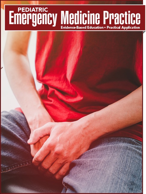
Pediatric Orthopedic Injuries: Evidence-Based Management in the Emergency Department - Calculated Decisions - Trauma CME
The Ottawa Ankle Rule shows the areas of tenderness to be evaluated in ankle trauma patients to determine the need for imaging.The Ottawa Knee Rule describes criteria for knee trauma patients who are at low risk for clinically significant fracture and do not warrant knee imaging.
The Ottawa Ankle Rule was derived to aid in the efficient use of radiography in the acute ankle and midfoot injuries.
It has been prospectively validated on multiple occasions in different populations and in both children and adults.
Sensitivities for the Ottawa Ankle Rule range from the high 90% to 100% range for “clinically significant” ankle and midfoot fractures. This is defined as a fracture or an avulsion > 3 mm.
Specificities for the Ottawa Ankle Rule are approximately 41% for the ankle and 79% for the foot, although the rule is not designed or intended to make a specific diagnosis.
The Ottawa Ankle Rule is useful in ruling out fracture (high sensitivity), but does poorly at ruling in fractures (many false positives).
Advice
Tips from the creators at the University of Ottawa:
Palpate the entire distal 6 cm of the fibula and tibia.
Do not overlook the importance of medial malleolar tenderness.
“Bearing weight” counts even if the patient limps.
Use with caution in patients aged < 18 years.
Precautions from the creators at the University of Ottawa:
Clinical judgment should prevail if the examination is unreliable due to:
» Intoxication
» Uncooperative patient
» Distracting painful injuries
» Diminished sensation in legs
» Gross swelling that prevents palpation of malleolar tenderness
Always provide written instructions.
Encourage followup in 5 to 7 days if pain and ability to walk do not improve.
Why and When to Use, Next Steps and Management
Why to Use
Patients who do not have criteria for imaging according to the Ottawa Ankle Rule are highly unlikely to have a clinically significant fracture and do not need plain radiographs. As a result, application of the Ottawa Ankle Rule can reduce the number of unnecessary radiographs by as much as 25% to 30%, improving patient flow in the emergency department (ED).
When to Use
The Ottawa Ankle Rule should be applied to all patients aged ≥ 2 years who have ankle or midfoot pain and/or tenderness in the setting of trauma.
An ankle x-ray series is only required if the patient has pain in the malleolar zone AND any of these findings:
» Bone tenderness at the posterior edge or tip of the lateral malleolus, OR
» Bone tenderness at the posterior edge or top of the medial malleolus, OR
» Inability to bear weight both immediately after injury and in the ED.
A foot x-ray series is only required if the patient has pain in the midfoot zone AND any of these findings:
» Bone tenderness at the base of the fifth metatarsal, OR
» Bone tenderness at the navicular, OR
» Inability to bear weight both immediately after injury and in the ED.
Next Steps
If ankle pain is present and there is tenderness over the posterior 6 cm of the tibia or fibula or the tip of the posterior or lateral malleolus, then an ankle-ray is indicated.
If the midfoot pain is present and there is tenderness over the navicular or the base of the fifth metatarsal, then a foot x-ray is indicated.
If there is ankle or midfoot pain and the patient is unable to take 4 steps both immediately after the injury and in the ED, then x-ray of the painful area is indicated.
Management
X-ray
RICE plan (Rest, Ice, Compression, Elevation)
Splinting/crutches and pain medication, pending outcome
Calculator Review Author
Calvin Hwang, MD
Clinical Assistant Professor
Department of Orthopaedic Surgery
Stanford University School of Medicine, Stanford, CA
Critical Actions
Patients who fulfill none of the Ottawa Ankle Rule criteria do not need an ankle or foot x-ray. Patients who fulfill either the foot or ankle criteria need an xray of the respective body part. Many experts would consider this score “one directional.” Because the rule is sensitive and not specific, it provides a clear guide of which patients do not need x-ray if all criteria are met; however, if a patient fails the criteria, the need for x-ray can be left to clinical judgment.
Evidence Appraisal
The original derivation study in 1992 included nonpregnant patients aged > 18 years who presented to Ottawa civic and general hospitals with a new injury < 10 days old. The initial pilot study included 155 patients, while the full-scale study included 750 patients. Any fracture that was not an avulsion of ≤ 3 mm was considered a clinically significant fracture. This resulted in the initial criteria: aged ≥ 55 years, inability to bear weight immediately after the injury and for 4 steps in the emergency department, or bone tenderness at the posterior edge or tip of either malleolus for the ankle. For the foot, criteria included pain in the midfoot and bone tenderness at the navicular bone, cuboid, or the base of the fifth metatarsal.
Further validation and refinement was completed in 1993, through a prospective study of 1032 patients in the validation and refinement phase of the study with 121 clinically significant fractures. The rules were further refined by removing the age cutoff from the ankle rule and cuboid tenderness from the foot rule, but the weight-bearing criterion was added to the foot rule. Sensitivity of the refined rule for both foot and ankle fractures was 100%, and ankle specificity increased to 41% and foot specificity to 79%.
An additional 453 patients were then prospectively enrolled in the second phase of the study, where the refined rules were validated, yielding a sensitivity of 100% for both ankle and midfoot fractures.
A study of 670 children aged 2 to 16 years at 2 separate sites found that the Ottawa Ankle Rule again had a sensitivity of 100% for both clinically significant ankle and midfoot fractures. This study also found that ankle x-rays could be reduced by 16% and foot x-rays by 29% if the rules were in use at the time of the study. Subsequent meta-analysis of the Ottawa Ankle Rule in children found 12 studies with 3130 patients and 671 fractures, with a pooled sensitivity of 98.5% and an overall reduction in x-ray utilization by 24.8%.
The Ottawa Knee Rule describes criteria for knee trauma patients who are at low risk for clinically significant fracture and do not warrant knee imaging.
The Ottawa Knee Rule was derived to aid in the efficient use of radiography in acute knee injuries.
It has been prospectively validated on multiple occasions in different populations and in both children and adults.
Numerous studies found sensitivities for the Ottawa Knee Rule of 98% to 100% for clinically significant knee fractures. One study did find a sensitivity of just 86%.
Specificities for the Ottawa Knee Rule typically range from 19% to 50%, although the rule is not designed or intended for specific diagnosis.
When the Ottawa Knee Rule is used appropriately, the number of knee x-rays obtained can be reduced by 20% to 30%.
The Ottawa Knee Rule is useful in ruling out fracture when negative (high sensitivity), but does poorly at ruling in fractures (many false positives).
Advice
Tips from the creators at the University of Ottawa:
Tenderness of the patella is significant only if it is an isolated finding.
Use only for injuries with a duration of < 7 days.
“Bearing weight” counts even if the patient limps.
Precautions from the creators at the University of Ottawa:
Clinical judgment should prevail if the examination is unreliable due to:
» Intoxication
» Uncooperative patient
» Distracting painful injuries
» Diminished sensation in legs
Always provide written instructions.
Encourage followup in 5 to 7 days if pain and ability to walk do not improve.
Why and When to Use, and Next Steps
Why to Use
Knee trauma patients who do not have criteria for imaging according to the Ottawa Knee Rule are highly unlikely to have a clinically significant fracture and do not need plain radiographs. As a result, application of the Ottawa Knee Rule can cut down on the number of unnecessary radiographs by 20% to 30%. This has proven to be cost-effective for patients without reducing quality of care (Nichol 1999).
When to Use
The Ottawa Knee Rule should be applied to all patients aged > 2 years who have knee pain and/or tenderness in the setting of trauma.
A knee x-ray series is only required for knee injury patients with any of these findings:
» Age ≥ 55 years, OR
» Isolated tenderness of the patella (with no bone tenderness of the knee other than the patella), OR » Tenderness of the head of the fibula, OR
» Tenderness of the head of the fibula, OR
» Inability to flex to 90°, OR
» Inability to bear weight both immediately after the injury and in the emergency department for 4 steps (unable to transfer weight twice onto each lower limb), regardless of limping.
Next Steps
Patients who do not have any of the Ottawa Knee Rule criteria present do not need an x-ray.
If 1 or more of the conditions are met, then an x-ray is recommended.
For significant nonbony injuries, often crutches and a knee immobilizer can be helpful to assist with ambulation.
Calculator Review Author
Calvin Hwang, MD
Clinical Assistant Professor
Department of Orthopaedic Surgery
Stanford University School of Medicine, Stanford, CA
Critical Actions
Patients who do not have any of the Ottawa Knee Rule criteria present do not need an x-ray. If 1 or more of the conditions are met, then an x-ray is recommended.
Many experts would consider this score “one directional.” Because the rule is sensitive and not specific, it provides a clear guide of which patients do not need x-ray if all criteria are met; however, if a patient fails the criteria, the need for x-ray can be left to clinical judgment.
Evidence Appraisal
The original derivation study by Stiell et al was done in 1995 and included non-pregnant patients aged > 18 years who presented to Ottawa civic and general hospitals with a new injury < 7 days old as a result of acute blunt trauma to the knee. The study enrolled 1054 subjects, of whom 68 had fractures, with 66 of them deemed to be clinically significant (not a simple avulsion fragment of < 5 mm in breadth without associated complete tendon or ligament disruption). Using recursive-partitioning techniques, the authors derived the 5 variables of their decision rule. If applied to the study population, their decision rule had sensitivity of 100% and specificity of 54% for identifying fractures and would lead to a 28% relative reduction in x-ray utilization.
Stiell et al then prospectively validated their decision rule in the same patient population. They performed telephone followup 14 days after the emergency department visit to determine the possibility of a missed fracture. Sensitivity of the decision rule was again 100%, identifying 63 clinically important fractures out of 1096 patients. Specificity was similar to the derivation study at 49%, and there was a 28% relative reduction in x-ray utilization.
Stiell et al prospectively implemented the decision rule in different teaching and community emergency departments. They found a relative reduction in x-ray usage of 26.4%, while maintaining a sensitivity of 100% for detecting 58 knee fractures out of 3907 patients, and a specificity of 48%. Moreover, there was a significant reduction in time to discharge and total medical charges in patients who did not get an x-ray.
The Ottawa Knee Rule has also been prospectively validated in populations outside of Canada. Two studies, 1 done in Spain and another in the United States, found that the Ottawa Knee Rule had a sensitivity of 100% and 98%, specificity of 52% and 19%, and a reduction in x-ray usage by 49% and 17%.
The rule was applied to children aged 2 to 16 years in a prospective, multicenter validation study in 2003. That study found the decision rule to be 100% sensitive in finding 70 fractures out of 750 children, with a specificity of 42.8% and a potential reduction in x-ray usage by 31.2%.
The Ottawa Knee Rule has also been compared to the Pittsburgh Decision Rule, another wellvalidated clinical decision rule. A cross-sectional comparison of the 2 rules showed that both had sensitivities of 86%, although the Pittsburgh Decision Rule was significantly more specific. However, this study only included patients aged 18 to 79 years and excluded pediatric patients.
Explain current treatment recommendations for pediatric orthopedic emergencies.
Physician CME Information
Date of Original Release: September 1, 2017. Date of most recent review: August 15, 2017. Termination date: September 1, 2020.
Accreditation: EB Medicine is accredited by the Accreditation Council for Continuing Medical Education (ACCME) to provide continuing medical education for physicians. This activity has been planned and implemented in accordance with the accreditation requirements and policies of the ACCME.
Credit Designation: EB Medicine designates this enduring material for a maximum of 4 AMA PRA Category 1 CreditsTM. Physicians should claim only the credit commensurate with the extent of their participation in the activity.
ACEP Accreditation: Pediatric Emergency Medicine Practice is also approved by the American College of Emergency Physicians for 48 hours of ACEP Category I credit per annual subscription.
AAP Accreditation: This continuing medical education activity has been reviewed by the American Academy of Pediatrics and is acceptable for a maximum of 48 AAP credits per year. These credits can be applied toward the AAP CME/CPD Award available to Fellows and Candidate Fellows of the American Academy of Pediatrics.
AOA Accreditation: Pediatric Emergency Medicine Practice is eligible for up to 48 American Osteopathic Association Category 2A or 2B credit hours per year.
Other Specialty CME: Included as part of the 4 hours, this CME activity is eligible for 4 Trauma CME credits, subject to your state and institutional requirements.
Needs Assessment: The need for this educational activity was determined by a survey of medical staff, including the editorial board of this publication; review of morbidity and mortality data from the CDC, AHA, NCHS, and ACEP; and evaluation of prior activities for emergency physicians.
Target Audience: This enduring material is designed for emergency medicine physicians, physician assistants, nurse practitioners, and residents.
Goals: Upon completion of this activity, you should be able to: (1) demonstrate medical decision-making based on the strongest clinical evidence; (2) cost-effectively diagnose and treat the most critical ED presentations; and (3) describe the most common medicolegal pitfalls for each topic covered.
Discussion of Investigational Information: As part of the journal, faculty may be presenting investigational information about pharmaceutical products that is outside Food and Drug Administration approved labeling. Information presented as part of this activity is intended solely as continuing medical education and is not intended to promote off-label use of any pharmaceutical product.
Faculty Disclosure: It is the policy of EB Medicine to ensure objectivity, balance, independence, transparency, and scientific rigor in all CME-sponsored educational activities. All faculty participating in the planning or implementation of a sponsored activity are expected to disclose to the audience any relevant financial relationships and to assist in resolving any conflict of interest that may arise from the relationship. Presenters must also make a meaningful disclosure to the audience of their discussions of unlabeled or unapproved drugs or devices. In compliance with all ACCME Essentials, Standards, and Guidelines, all faculty for this CME activity were asked to complete a full disclosure statement. The information received is as follows: Dr. Lien, Dr. Sanders, Dr. Claudius, Dr. Mishler, and their related parties report no significant financial interest or other relationship with the manufacturer(s) of any commercial product(s) discussed in this educational presentation. Dr. Lee made the following disclosure: royalty payments, UpToDate. Dr. Jagoda made the following disclosures: Consultant, Daiichi Sankyo Inc; Consultant, Pfizer Inc; Consultant, Banyan Biomarkers Inc; Consulting fees, EB Medicine.
Commercial Support: This issue of Pediatric Emergency Medicine Practice did not receive any commercial support.
Earning Credit: Two Convenient Methods: (1) Go online to www.ebmedicine.net/CME and click on the title of this article. (2) Mail or fax the CME Answer And Evaluation Form with your June and December issues to Pediatric Emergency Medicine Practice.
Hardware/Software Requirements: You will need a Macintosh or PC with internet capabilities to access the website.
Additional Policies: For additional policies, including our statement of conflict of interest, source of funding, statement of informed consent, and statement of human and animal rights, visit https://www.ebmedicine.net/policies.