
|
|
This issue of Emergency Medicine Practice provides an overview of thoracic imaging modalities and guidance on the indications for each test in emergency practice.
You return to the ED at 7am to find the night doc repairing a complex upper extremity laceration. Evidently,it was a hectic night, but your colleague insists on finishing the repair he started three hours earlier. Sign out will wait. There are three charts in your rack. You scan the triage notes:
"32-year-old man with asthma exacerbation, dyspnea, and chest pain- albuterol nebs and prednisone ordered." Next, "28-year-old female with chest pain and dyspnea." Third, "62-year-old male with upper back pain." His ECG is non diagnostic but the blood pressure of 200/120 piques your interest and guides you to his room. This hypertensive one hour prior to arrival; when he describes the pain as ripping, you consider an aortic emergency...
A request for an analgesia order prompts you to see the 28-year-old female next. She report abrupt onset of severe pleuritic left chest pain while breast feeding her 4-day-old daughter delivered by c- section. The pain has been constant since it began two hours ago and is accompanied by dyspnea and non productive cough. She appears tachypneic and apprehensive. The lungs are clear. She attributes mild legs swelling to pregnancy...
From behind a curtain you hear the asthmatic insisting he has to leave for work. He's a 32-year-old male "frequent-flier" asthmatic well known to the ED staff. Seeing his face, you realize you've intubated him before but the appears quite comfortable as he starts his third neb. He'd been battling this exacerbation all night but called 911 when his inhaler ran dry. The chest tightness referred to an the triage note resolved with the first treatment. He denies any infectious symptoms. The lung exam reveals equal breath sounds with good air exchange and minimal expiratory wheezing. The nurse asks if you want a chest x-ray...
Just as you think you have the department under control, you are notified that the clinic is sending a 44-year-old woman with shortness of breath. She has a history of cocaine use, myocardial infarction, and frequent heart failure. No sooner than when you hang up, the patient arrives propped upright on the stretcher appearing anxious, uncomfortable, and tachypneic. You note normal neck veins and the lung sounds are clear, but markedly diminished on the right. Listening again, you think maybe this isn't just another episode of acute decompensated heart failure...
AP - Antero-Posterior
CT - Computed Tomography
CXR - Chest Radiograph
EFAST - Extended Focused Abdominal Sonogram for Trauma
ECG - Electrocardiogram
ECHO - Echocardiography
FAST - Focused Abdominal Sonogram for Trauma HF - Heart Failure
LBBB - Left Bundle Branch Block
LVH - Left Ventricular Hypertrophy
MRI - Magnetic Resonance Imaging
PA - Posteroanterior
SPECT - Single Photon Emission Computed Tomography
TEE - Transesophageal
TTE - Transthoracic
V/Q - Ventilation / Perfusion
The selection of thoracic imaging studies ideally should be based on carefully designed studies which determine the sensitivity, specificity, and positive and negative predictive values of the test. Many times, such specific data is not available and clinicians must base their choice on local practice or non evidence-based recommendations from books or other publications. One of the goals of this article is to establish recommendations available in the literature and to place them in the context of the evidence from which they were derived. This article also hopes to identify areas in need of further study.
Thoracic imaging studies range from those used routinely and frequently, such as the chest radiograph, to those that the practicing emergency physician will probably consider relatively infrequently and perhaps only in highly acute situations, such as imaging for aortic dissection. The chest radiograph is so ubiquitous that it is often ordered routinely without much consideration for the indications. For example, the portable anteroposterior chest radiograph is, along with a lateral cervical spine film and an anteroposterior pelvis, part of the basic screening radiology evaluation of the trauma patient, as recommended by the American College of Surgeons Advanced Trauma Life Support Course (ATLS).1 While this recommendation is appropriate for the major trauma patient, its application to many patients with far less than major, multi-system trauma as part of the "cookbook" approach to the trauma patient, has unquestionably led to countless unnecessary studies.
The ATLS recommendations are voluminous and detailed. However, this particular set of expert consensus guidelines does not specify the methodology used to create the recommendations and does not specify the strength of the evidence upon which they are based. Therefore, the user cannot easily differentiate between strong recommendations based on prospective studies using a gold standard, and weak recommendations based on case studies and anecdotal experience. While each chapter of the ATLS text contains a compendium of references, recommendations are not specifically referenced to the source(s). 1
The American College of Radiology has developed a novel set of guidelines for the use of imaging studies and has published these as "ACR Appropriateness Criteria."2-8 Each guideline was developed by a panel of experts and begins with a summary and critique of the literature. This review is then used to assess the appropriateness of individual imaging studies for various clinical situations and the appropriateness is rated on a scale of one to nine, with one being the least appropriate and nine being the most appropriate. For example, for adults less than 65 years of age with possible rib fracture, rib films are given an appropriateness rating of two, while the chest radiograph is given an appropriateness rating of eight.2
The indications for chest radiography in patients of various ages with respiratory symptoms have been outlined in guidelines from the American College of Radiology, the American Thoracic Society, and the American College of Emergency Physicians.3,-6,8-10 In addition, there are a number of prospective studies covering this topic.11-15Similarly, the American College of Radiology and a number of prospective studies have verified the lack of utility of the chest radiograph in acute asthma exacerbations.4,16-19However, this conclusion cannot be expanded to patients with chronic obstructive pulmonary disease, where chest radiograph has been shown to have a much higher level of appropriateness.4,20
The use of ultrasonography in the evaluation for thoracic pathology is a rapidly developing field. Prospective studies to determine the sensitivity of ultrasound for various clinical problems have been published, but, in most cases, results have to be considered preliminary due to the limited number of confirmatory studies. This remains an area in need of study and an opportunity for emergency medicine researchers to perform well-designed prospective studies and meaningfully add to the emergency medicine literature.
Similarly, with the advent of multi-row detectors, the use of computed tomography (CT) scanning of the chest has greatly expanded. As a result of the technological advances, the sensitivity and specificity of the test for various indications have increased. Strong, validated evidence shows that CT is now the modality of choice for the diagnosis of mediastinal hemorrhage, aortic trauma, aortic dissection, and aortic aneurysm. 21,22
The evaluation of the patient with dyspnea and potential pulmonary embolism has also undergone marked change. This has been an area of intense study with many robust studies of the use of CT scanning. 23-33There are large, well-designed, prospective studies in this area, as well as meta-analyses and thoughtful editorials to assist in assimilating this data.34-39
The use of imaging in patients with possible acute coronary syndromes is also undergoing rapid change. The American College of Cardiology (ACC), the American Heart Association (AHA), and the American Society of Echocardiography have issued joint guidelines for the clinical use of echocardiography.40 With the American Society for Nuclear Cardiology, the ACC and AHA have issued joint guidelines for the use of nuclear medicine scanning in the diagnosis of acute coronary syndromes.41Recently, multi-detector computed tomography (MDCT) for non-invasive coronary angiography has received attention in regard to diagnosing acute coronary syndromes.42-48 However, a recently published report of a large multicenter trial found a high rate of unevaluable segments, leading to questions regarding the clinical role of MDCT in evaluating coronary artery stenosis.49
In choosing an imaging modality, the emergency physician attempts to optimize diagnostic accuracy, rapidity of testing, patient safety, and expense. Prudent medical decision making begins with a deliberate history and physical examination yielding a reasonable differential diagnosis. The list of pathology to be excluded directs what imaging is pursued.
The chest radiograph (CXR) serves as a fast initial study for most thoracic complaints. The CXR exposes the patient to minimal radiation and does not necessarily require travel out of the ED. These advantages can contribute to overuse however.
The ventilation/perfusion (V/Q) scan was once the prevailing study for the evaluating pulmonary embolism, but the pulmonary scintigram has been widely supplanted by MDCT. Limitations of the V/Q scan include radiation exposure and handling radio nuclides, lengthy image acquisition time, transport of patients to areas where close monitoring is challenging, and frequent difficulty in study interpretation, especially in patients without a normal chest x-ray.23
MDCT provides excellent detail of the aorta and pulmonary vasculature with scans acquired in a single breath-hold.21 MDCT may reveal alternate diagnoses. As new ED designs incorporate CT scanners within the department, transport times and risks of decompensation away from the ED are minimized. Risks of IV contrast administration include renal impairment and allergic reaction, though these effects have been mitigated by newer generations of ionic contrast agents.
Echocardiography (ECHO) is particularly useful in patients too unstable to leave the resuscitation room. ECHO may detect right ventricular dilitation suggestive of pulmonary embolism or confirm aortic dissection without radiation. Diagnostic accuracy is operator dependent.50
Magnetic Resonance Imaging (MRI) provides excellent detail of thoracic pathology without radiation. This use in ED patients is often limited by prolonged image acquisition, distance from the clinical area, and expense.51,52 Another limitation of MRI is that it can not be used in patients with implaned devices such as pacemakers and automatic inplantable cardioverter defibrillators.
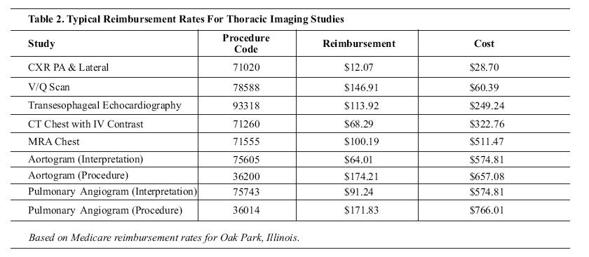
Emergency physicians face both continued emphasis on cost containment and the introduction of newer, often more expensive, imaging options. An evidence-based, cost-effectiveness analysis directs rational utilization of limited medical resources. Although charges or reimbursement may serve as proxies for cost, they often do not accurately reflect the resources consumed; see Table 2. Though not easily quantified, hidden imaging costs include ED staff utilization, opportunity costs, and risks associated with the procedure. Travel from the ED for imaging requires staff to deliver and collect the patient and may occupy a nurse for continuous monitoring. Longer image acquisition and radiologist interpretation times delay patient disposition. This translates into the cost of forgone opportunity to direct resources toward the next patient in need.
The risk of exposure to ionizing radiation is also an imaging cost. The expanded use of computed tomography, with its associated increase in radiation exposure compared to plain radiography, has led to renewed concerns about the total dose of ionizing radiation and the potential for increased rates of cancer, which can occur soon after exposure or up to decades later. This concern is especially high for the pediatric population because their post-exposure life span is greater and they have a higher number of dividing cells than adults.53CT scanning is estimated to account for about 10% of diagnostic radiology examinations, but is responsible for up to two-thirds of the total radiation dose delivered to the population.54 There is no current consensus on whether there is such a thing as a "safe dose" or what constitutes a reasonable exposure threshold.53 While specific guidelines have not been published, the International Commission on Radiological Protection is expected to publish guidelines in 2007 which are anticipated to include recommendations for limiting exposure via medical x-ray sources.55The awareness level concerning radiation dose and possible risks associated with CT scans is low among radiologists (47%), emergency physicians (9%), and patients (3%), based on results of a survey by Lee et al published in 2004.56
Another concern when considering imaging techniques is the risk for development of contrast-induced nephropathy, which is defined as the elevation of serum creatinine more than 0.5 mg/dL within three days of contrast media administration. Numerous risk-reduction strategies have been investigated. Adequate intravenous volume expansion with isotonic crystalloid, beginning 3 to 12 hours before the procedure and continuing for 6 to 24 hours afterward, can lessen the probability in high risk patients. It is not known whether oral hydration is effective. According to the Contrast-Induced Nephropathy Working Panel, of the pharmacologic agents that have been suggested, theophylline, statins, ascorbic acid, and prostaglandin E1 deserve further investigation. N-acetylcysteine has not been shown to be consistently effective. Diuretics are considered to be potentially detrimental. Nephrotoxic drugs, such as non-steroidal antiinflammatory agents and aminoglycosides, should be withdrawn before contrast administration.57
Another approach to the reduction of the risk for development of contrast-induced nephropathy is through the use of an isosmolar contrast medium, such as iodixanol (Visipaque). A recently published metaanalysis of 16 double-blind studies including 2727 patients found that iodixanol was associated with smaller rises in serum creatinine and lower rates of contrast-induced nephropathy than low-osmolar contrast media.58
Obtaining images must never take precedence over clinical evaluation and continuous monitoring of potentially unstable patients. Whenever possible, imaging should be accomplished at the bedside for unstable patients. Portable chest radiography and bedside ultrasound often provide valuable information without compromising care. However, many patients will subsequently require more intensive imaging studies that will involve the transportation of the patient outside of the ED proper.
Up-to-date planning and designing of facilities can help to limit distance and time outside the ED. Current use of computed tomography for the acute evaluation of patients is so extensive that it makes very good sense to have the CT facility in or adjacent to the ED. As the use of MRI expands and more acute indications are explored, the location of MRI units in close proximity to the ED will also become more advantageous.
The most commonly ordered imaging study of the thorax remains the chest radiograph (CXR) with routine studies including the posteroanterior (PA) and lateral views. Patients who cannot be transported to the radiology suite are often studied using a portable anteroposterior (AP) view.
General guidelines for the interpretation of chest radiographs have been well-outlined by Schwartz et al;59,60 see Tables 3 and 4.
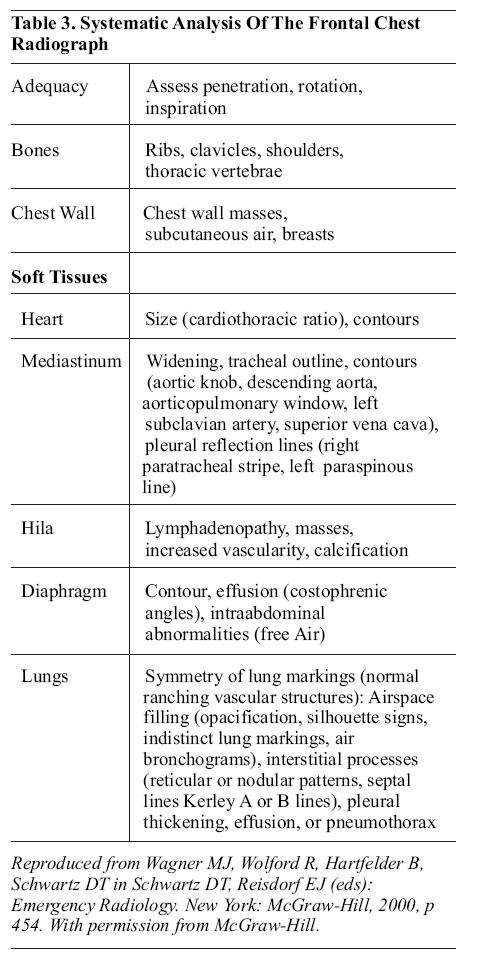
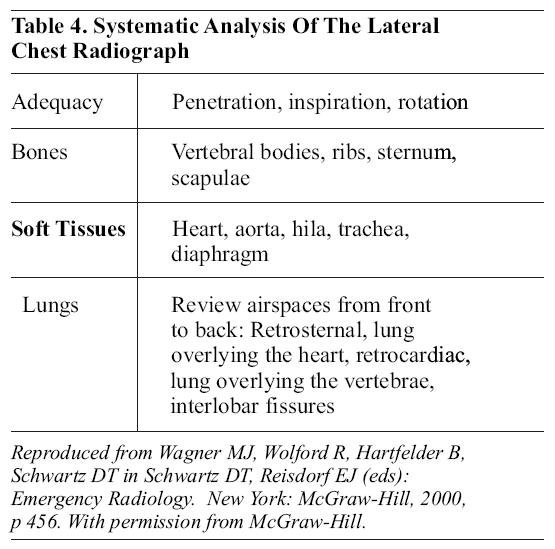
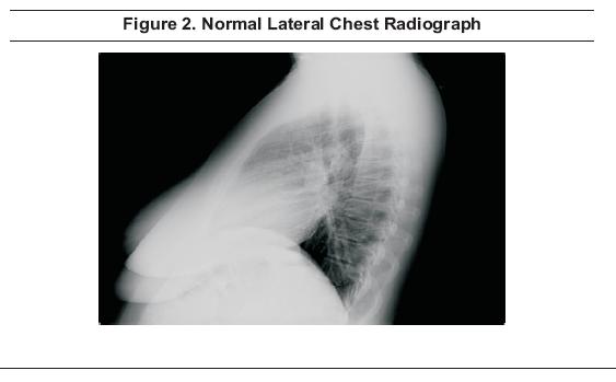
In the interpretation of the lateral PA and AP chest (Figures 1 and 2 ), the first concern is to assure the adequacy of the film. The entire thorax should be seen, including the apices, lateral chest walls, entire diaphragm, and both costophrenic angles.

Penetration should be such that the lower thoracic vertebral bodies are faintly visible behind the heart and the image should be positioned so that the mid-point between the clavicular heads is superimposed over the spinous processes of the thoracic vertebrae. The film should be shot in inspiration so that the right costophrenic sulcus is below the posterior costovertebral junction of the 10th rib.
Systematic analysis of the images begins with the assessment of the bony thorax, including the ribs, clavicles, shoulders, and thoracic vertebral bodies.
Heart size is considered normal if the cardiothoracic ratio is less than 0.5; cardiomegaly is present if the cardiothoracic ratio is greater than 0.5. Thoracic width is measured at the widest point, i.e., the lung base. The cardiac contours are assessed for evidence of chamber enlargement.
Mediastinal widening is present when the mediastinum measures greater than 8 cm at the aortic arch in adults or the mediastinum:chest width ratio is greater than 0.25 in children. The trachea should be in the midline. The contours of the mediastinum are assessed, noting the aortic knob, descending aorta, aorticopulmonary window, left subclavian artery, superior vena cava, right paratracheal stripe, and left paraspinous line.
The hila of the lungs are assessed looking for adenopathy, masses, vascularity, and calcifications.
The diaphragm is evaluated looking at the contour, costophrenic sulci, and for any evidence of free air in the space beneath the diaphragm.
The lungs are evaluated for symmetry. Lung markings should be visible as a branching vascular pattern. The air spaces are evaluated for evidence of opacification, silhouette signs, or air bronchograms. The retrosternal space, retrocardiac space, and interlobar fissures are assessed. Interstitial processes may be detected as a reticular pattern, a nodular pattern, or as septal lines (Kerley A or B lines).
The pleura may show thickening or the pleural space may contain fluid (effusion) or air (pneumothorax).
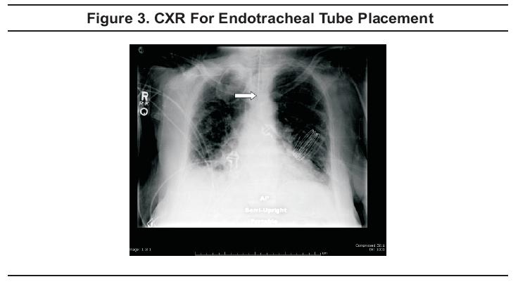
The CXR is an essential part of the assessment of endotracheal tube placement (Figure 3). The preferred location is 3 to 4 cm above the carina. In addition, the CXR may exclude pneumothorax and can potentially confirm various diagnoses, such as pneumonia or congestive heart failure.61
Ultrasound guidance of central line placement may decrease the time required and the number of attempts necessary prior to successful cannulation.62However, while it has the potential to improve successful line placement and to minimize complications, such a reduction in complication rate has yet to be confirmed.63,64
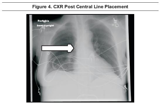
A chest radiograph has long been recommended following any attempt at placement of a cervical or thoracic central line. CXR allows for the assessment of the location of the tip of the catheter (Figure 4) and assists in ruling out pneumothorax or hemothorax, although supine films are of limited value in assessing for pneumothorax or hemothorax due to anterior layering of air and/or posterior layering of blood in these views. However, confirmatory radiographs may not be needed after straight-forward placements.65
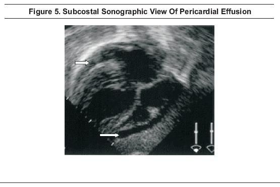
The portable AP chest radiograph, along with a lateral cervical spine film and AP pelvis, remains a part of the screening radiology evaluation for the blunt multisystem trauma patient, as recommended by the American College of Surgeons Advanced Trauma Life Support Course.1In addition, the focused abdominal sonogram for trauma (FAST) is recommended not only for the evaluation of potential intra-abdominal injury but for the evaluation of the pericardial sac (Figure 5).1,66-68
In addition to the sub-costal view of the heart and pericardium, standard views include the right upper quadrant, the left upper quadrant, and the pelvis. Views of the right and left paracolic gutters are often added and it may be possible to visualize blood superior to the hyperechoic diaphragm in the presence of hemothoraces. While the FAST exam is relatively reliable in detecting free intraperitoneal blood, it has limited utility in detecting solid organ injury or retroperitoneal bleeding.
Ultrasound has been shown to be more sensitive than supine AP chest radiograph for the detection of traumatic pneumothoraces69 and, in some centers, thoracic ultrasound is performed routinely along with the traditional FAST scan, creating the extended focused abdominal sonogram for trauma (EFAST).70
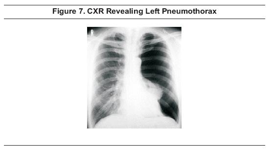
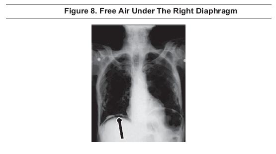
In the setting of penetrating trauma, no imaging is required if the patient is hemodynamically unstableand does not respond to resuscitation with crystalloids and blood. Operative intervention is required in these cases. In stable patients with penetration above the umbilicus or for those with suspected thoracoabdominal injury, an upright CXR is the most commonly used study to evaluate for the presence of hemothorax (Figure 6), pneumothorax, (Figure 7), or intraperitoneal air (Figure 8). Serial CXR's may be used when suspicion is high and initial screening radiographs are negative.1 Using the supine AP CXR, the presence of subcutaneous emphysema or the radiographic deep sulcus sign may be useful in diagnosing small pneumothoraces. The deep sulcus sign is detected by noting lucency and a sharp, angular appearance of the costophrenic angle on the involved side.71
However, some studies showed that ultrasound is more sensitive than the CXR in diagnosing pneumothoraces69 and hemothoraces.8 An ultrasonic deep sulcus sign may be noted sonographically.72
As with blunt trauma, the FAST scan is used to evaluate the pericardial sac (Figure 5) and to assess for blood in Morison's pouch (the hepatorenal interface), the splenorenal interface, and in the pelvic spaces; the EFAST scan can be used to evaluate the thorax simultaneously. 1,70
MDCT scan is superior to supine chest radiographs in diagnosing pulmonary contusion. On CT, contusions appear as patchy or diffuse air space filling that tends to be peripheral, nonsegmental, and geographic in distribution. CT is also the imaging study of choice for transmediastinal gunshot wounds, since CT is able to visualize wounds that penetrate the great vessels, pericardium, esophagus, trachea, and thoracic spine. CT is less expensive, less time-consuming, and less invasive than angiography or endoscopy and these tests can generally be avoided if MDCT confirms that the wound track does not come in close proximity to these structures.73
Rib views have traditionally been used for detection of rib fractures in patients who have been subjected to direct blows or compressive injuries to the chest, but often add little to the management of the patient. The ACR rates specialized rib views as having a low level of appropriateness (2/9) for adults less than 65 years of age.2While not specifically indicated, the ACR rates these views at a moderate level of appropriateness (5/9) for adults greater than 65 years of age.
The ACR recommends rib views as more appropriate for children under 14 years of age (8/9) since children have more compliant rib cages and the presence of fracture(s) is associated with significant trauma and increased associated injury. In children less than three years of age, rib fractures are frequently a marker of abuse.74
The chest radiograph is appropriate at any age (8 to 9/9) when the diagnosis of rib fracture is under consideration, and is primarily used to rule out associated pulmonary injury.2
In the past, the literature has stressed the importance of rib fractures, especially those of the first and second ribs, as predictors of aortic injury. However, several studies have demonstrated no increased likelihood of aortic injury with upper rib fractures.2,75 No additional imaging studies are mandated by these findings alone.
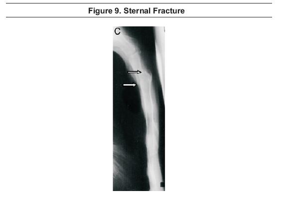
Sternal fractures (Figure 9) are reported to be associated with severe injuries. In a retrospective review of 200 sternal fractures, von Garrel et al reported injuries to the thoracic vasculature, including the heart, in approximately 30% of cases, and such injuries were increased with displacement of sternal fragments. Fatal heart injuries were frequently seen in conjunction with sternal fractures in patients who fell from significant heights. Spinal injuries were associated with sternal fractures in 13% of cases and were most likely in fractures with involvement of the manubriosternal joint.78
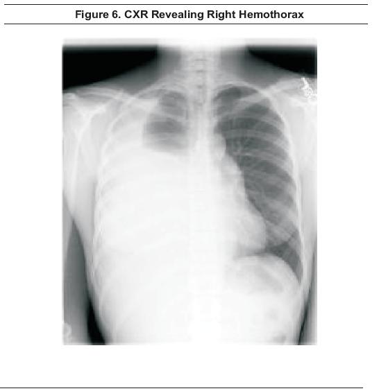
Hemothorax is most often detected by the finding of fluid in the pleural cavity on screening CXR (Figure 6). However, ultrasonography can detect hemothoraces not evident on CXR and is rapid and accurate; sensitivity of ultrasound is reported at 92%, specificity at 100%, positive predictive value at 100%, and negative predictive value at 98%.79, 80
Angiography plays a role in the evaluation of the patient with hemothorax and may identify occlusion, active hemorrhage, or pseudoaneurysm. A potential advantage of angiography is that, when specific bleeding sites are identified, one can proceed to selective embolization of the internal mammary or intercostal artery, which may be an effective alternative to thoracotomy.81,82 While this treatment modality is promising, the number of cases studied is small.
Further study is needed before firm recommendations can be made.
The primary modality currently used for detection of pneumothorax or pneumomediastinum is the CXR (Figure 7). Inspiratory and expiratory views probably do not improve the detection of pneumothoraces above the standard CXR.83,84,85 A prospective, randomized review of 178 patients paired inspiratory and expiratory chest radiographs with and without pneumothoraces; inspiratory and expiratory upright films were found to be equally sensitive for pneumothorax detection.84 Films must be perused carefully since small pneumothoraces can easily be missed and overlying skin folds can simulate pneumothoraces. Ultrasound is more sensitive than AP CXR for the detection of pneumothorax and demonstrates good agreement with CT scan.69
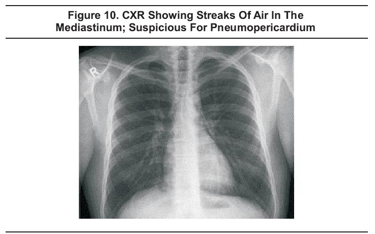
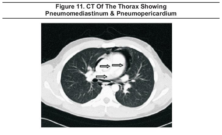
Pneumopericardiummay result from blunt chest trauma, pneumothorax, pneumoperitoneum, pneumomediastinum, tracheobronchial tears, or esophageal tears. It may be seen on CXR (Figure 10), but is best diagnosed using CT scan of the thorax and abdomen (Figure 11) which allows for the additional detection of concomitant injuries.86,87
Hemorrhage into the mediastinum is suspected when the supine AP CXR shows abnormal mediastinal contours (Figure 12). A number of radiographic findings have been promulgated as indicative of mediastinal hemorrhage, but, in a study comparing radiograph interpretation in normal patients and patients with mediastinal hemorrhage, Woodring found only five signs to be helpful; see Table 5.88 The aortic contour is considered to be abnormal when the aortic knob is enlarged, irregular, or indistinct. The mediastinum is considered to be widened on the supine AP CXR when the width is 8 cm or greater when measured just above the aortic knob. An apical cap is formed when blood dissects above the lung on either side; but a left apical cap is more indicative of mediastinal bleeding than one on the right. Displacement of a nasogastric tube to the right at the level of T4 is also suggestive of a mediastinal hematoma. The right paratracheal stripe is a space between the right tracheal wall and the adjacent lung and pleura. With hemorrhage into the mediastinum, this potential space can fill with blood and become distended. Based on a study of 102 consecutive patients using thoracic arteriograms as the gold standard, widening to greater than 5 mm is suggestive of mediastinal hemorrhage; a paratracheal stripe was reported to be associated with major arterial injury in 23% of cases.89

CT of the chest is 100% sensitive and 99.7% specific for mediastinal hemorrhage. The positive predictive value is 89% while the negative predictive value is 100%, giving an overall diagnostic accuracy of 99.7%.90 CT is accurate for the detection and localization of both hemomediastinum and direct signs of aortic injury, and has largely supplanted aortography for the diagnosis of these problems.90
Chest radiography is frequently used as an initial screening tool in patients with possible aortic injury, but there are no CXR findings with both high sensitivity and high specificity for aortic injury. A mediastinum greater than 8 cm at the level of aortic knob (Figure 12) has a sensitivity greater than 90% but a low specificity. Thoracic spine fracture, first rib fracture, rightward deviation of a nasogastric tube, depression of the left mainstem bronchus, and widened paraspinal line are all findings with specificity greater than 90% but low sensitivity, and no significant improvement in overall accuracy was achieved by combining radiographic findings.91False positive and false negative findings occur with each x-ray sign, and in 1 to 2% of cases, the supine AP CXR is normal in the presence of a great vessel injury.92
As follow up for an abnormal CXR, computed tomography of the chest has a sensitivity of 100% and specificity of 99.7%.90
When there is evidence of aortic injury on CT, either aortography or surgery is indicated. An aortogram is useful when there is evidence of mediastinal hematoma adjacent to the aorta, but no aortogram is required for negative CT or for hematomas not adjacent to the aorta.90
A retrospective review of 200 patients with chest trauma found that pulmonary contusion (Figure 13) was the most common thoracic injury.93

CT gives the ability to better define the extent of the injury. Contusions appear radiodense and are usually peripheral, nonsegmental and nonlobar. The increased lung density is due to distal lung hemorrhage and edema.94
In the setting of penetrating trauma to the heart or lung, evaluation for pericardial hemorrhage is best carried out by echocardiography. The best view is the subcostal view in which fluid or blood will appear as an anechoic area surrounding the heart (Figure 5). Fluid will collect posteriorly first. If seen only anteriorly, the finding may be due to fatty deposition. Other potentially useful views are the parasternal long axis, parasternal short axis, and apical views. Sensitivity is reported to be 100% and specificity is 96.9% to 100%.97,98
Cardiac tamponade is a cardiovascular emergency requiring rapid diagnosis.99Sonographic criteria for the diagnosis of tamponade include diastolic collapse of the right ventricle or right atrium, possible collapse of the left atrium and ventricle, and distended inferior vena cava without respiratory variation. Transthoracic drainage under ECHO guidance is the recommended treatment, and has largely replaced the standard "blind" subxiphoid approach to pericardiocentesis commonly employed in the past.100
In the setting of tracheobronchial injury, lateral neck films may show air in soft tissues. CXR may show pneumomediastinum or pneumothorax ( Figure 7 ).101 CT of the chest with 3-D reconstruction of the tracheobronchial tree may be equivalent or superior to bronchoscopy.102,103
Based on a retrospective review of 14 patients with esophageal perforation, Ghanem et al reported that the most common CXR finding was pleural effusion (64%), which was bilateral 60% of the time. When the effusion was unilateral, it was more commonly on the left. Pulmonary infiltrates were present in 64% of the cases and were most commonly bilateral. If unilateral, left-sided infiltrates were more common. Other CXR findings included pneumomediastinum (21%), pneumothorax (14%), and pneumopericardium (14%). Esophagography is indicated when an esophageal tear is suspected.86 The initial study should use water-soluble contrast medium, followed by a barium study if the water-soluble contrast study is negative. Positive findings on either study include extravasation (64%) and submucosal contrast medium collection (36%). Historically, endoscopy has been recommended if there is a high probability of injury and negative esophagography. However, CT has been shown to have sensitivity and specificity of 100% after suspected perforation.104CT findings of mediastinitis include increased attenuation of mediastinal fat (100%), pleural effusions (85%), free mediastinal gas bubbles (58%), localized mediastinal fluid collections (55%), sternal dehiscence (40%), mediastinal lymph nodes (35%), lung infiltrates (35%), pericardial effusion (28%), and pleuromediastinal fistula (3%).104
According to ATLS, CXR findings consistent with diaphragmatic injury include elevation or blurring of the diaphragm (Figure 14), hemothorax, abnormal gas shadow obscuring the hemidiaphragm, or gastric tube positioned in the chest.1

Findings on CT are similar. Based on the review of CT examinations of 179 patients with blunt trauma, Nchimi et al reported the following findings as strong predictors of blunt diaphragmatic rupture: Discontinuity, thickening, segmental non-recognition, intrathoracic hernia of abdominal viscera, elevation, hemothorax, and hemoperitoneum. Although not yet validated by other studies, the combination of discontinuity, thickening, and segmental nonrecognition was reported to be 100% sensitive.105 While CXR findings, especially displacement of a gastric tube, may be diagnostic of diaphragmatic injury, CT increases the accuracy of the diagnosis significantly.105
ACR's Appropriateness Criteria rates CXR as highly appropriate (8/9) for most patients with a complaint of dyspnea regardless of physical findings, other symptoms, or risk factors for cardiopulmonary disease.3 CXR may demonstrate pulmonary infiltrates, vascular congestion, pneumothorax, pleural effusions, or neoplastic disease. Indirect evidence of thromboembolic disease may also be seen. For those under the age of 40 with a negative physical examination, the appropriateness is described as being influenced by severity and duration of dyspnea and the presence of other symptoms or risk factors for cardiovascular, pulmonary, and neoplastic diseases.3 While CT is not recommended for the initial evaluation of patients with dyspnea, except for patients with suspected pulmonary embolism, the ACR rates CT as appropriate (8/9) at any age when clinical evaluation, plain films, and laboratory studies are non-diagnostic.3 Plain CT is useful for detecting many diseases that may present with dyspnea, such as emphysema, sarcoidosis, and lung cancer.
Ventilation/perfusion (V/Q) lung scanning has been the primary tool for imaging pulmonary embolism in the past. In the Prospective Investigation of Pulmonary Embolism Diagnosis (PIOPED) study, the sensitivity of a normal or near-normal V/Q scan was shown to be 99%, with specificity of 98%. However, 78% of scans were read as low or intermediate probability. In addition, the overwhelming majority of patients without pulmonary embolism still had abnormal scans.23 V/Q scans require two hours to perform. One advantage is that V/Q scans result in less exposure to ionizing radiation than CT scans so they may be considered more useful for pregnant patients and patients that cannot tolerate intravenous contrast due to hypersensitivity or renal insufficiency.23
CT angiography of the chest has several advantages over either V/Q scanning or pulmonary angiography in the evaluation of the patient with possible pulmonary embolism. It is faster than either V/Q scanning or angiography. It is more practical in dyspneic patients and requires less contrast than angiography. It is generally more available than V/Q scanning or angiography and it may detect other important diagnoses when pulmonary embolism is not present24.
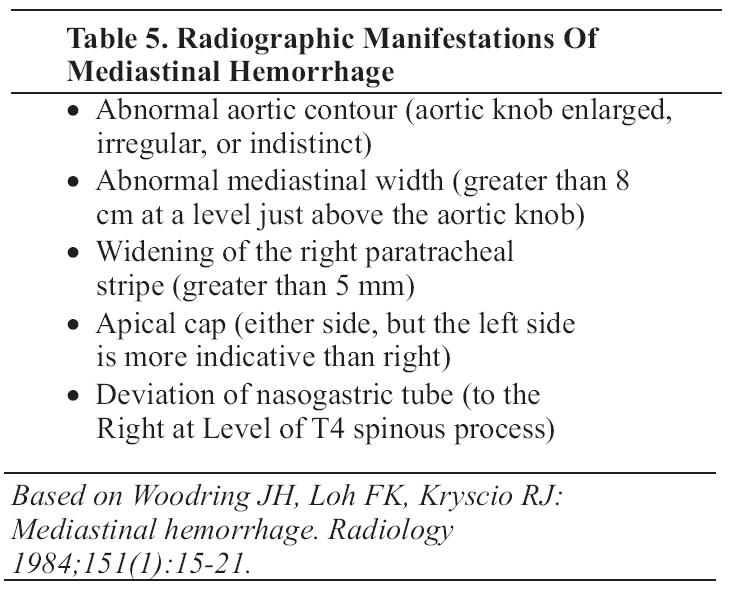
By 2001, CT scanning was being used more than lung scanning to investigate suspected pulmonary embolism.25Even with older generation scanners, CT could image from the main pulmonary arteries to the segmental and possibly sub-segmental arteries (Figure 15),24 but inter-observer agreement was poorer for sub-segmental arteries26.
With 16-slice multidetector-row CT scanners now commonly available, the entire chest can be imaged with excellent resolution, requiring a breath-hold of less than 10 seconds. These scanners can reliably diagnose tiny emboli in sub-segmental vessels.106 The clinical significance of sub-segmental emboli is unclear. In a study that included 67 patients with isolated subsegmental pulmonary emboli, Eyer et al reported that 37% did not receive anticoagulation and that there was no evidence of recurrent thromboembolism on follow up.107 Further study to confirm these findings is needed.
A positive CT result is an intraluminal filling defect or vascular occlusion24 (Figure 15). Reported sensitivities vary widely, being affected significantly by the generation of scanner used. While large series using specific generations of scanners are yet to be published, Russo et al published a meta-analysis of the relevant literature from 1995 to 2004. This review showed the sensitivity and specificity to have increased from 37 to 94% and from 81 to 100% respectively, primarily due to the possibility of depicting subsegmental clots.108
The PIOPED II trial was a prospective, multicenter investigation of the accuracy of multidetector CT angiography alone and combined with venous-phase imaging (CT angiography-CT venography) for the diagnosis of acute pulmonary embolism.38Combined CT angiography-CT venography was found to have higher diagnostic sensitivity than CT angiography alone, but the increased diagnostic yield is probably not enough to justify the additional radiation.39 The predictive value of either approach is high when the result is concordant with clinical assessment, but clinicians should be wary and consider additional testing when results are discordant with clinical probability.38,39
Echocardiography is not a sensitive test for pulmonary embolism. Sonographic criteria for pulmonary embolism include right ventricular dilation, septal wall motion abnormality, decreased right ventricular contractility, elevated pulmonary artery or right ventricular pressures, moderate to severe tricuspid regurgitation, and visualization of the clot in the right ventricle or pulmonary artery. Sensitivity is only 41% and specificity is 91%.33,110
Magnetic resonance imaging of the chest can be performed relatively rapidly, but continues to have limited availability. The diagnostic performance of MRI is similar to that for V/Q scanning. One advantage is that MRI does not use ionizing radiation and therefore may be safer for imaging pregnant patients.24
ACR's Appropriateness Criteria for CXR in uncomplicated asthma is only 4/9.4 A CXR is often recommended for the first episode of wheezing. Based on a retrospective review of 90 episodes of acute asthma in adults, Findley et al reported that the chest radiograph findings were most commonly normal (55%), hyperinflated (37%), or showed interstitial changes previously identified on radiographs (7%). Only one new alveolar infiltrate was found in this series (1%). They concluded that, in the setting of acute asthma, the chest radiograph is indicated only when pneumonia or pneumothorax is suspected.16 Abnormal CXR findings are more common in children with first episodes of wheezing (6 to 16%), but, in the absence of clinical variables, these findings rarely affect the acute management of the patient.17-19
Approximately one-fourth of radiographic abnormalities seen in patients with apparent exacerbations of chronic obstructive pulmonary disease are not predictable on the basis of high-risk criteria. Consequently, routine chest radiography should be considered.20 ACR's Appropriateness Criteria for uncomplicated COPD is 7/9; the appropriateness rating increases to 9/9 in the presence of leukocytosis, bandemia, chest pain, or cardiac history.4
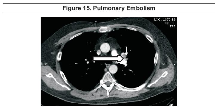
In the acute or exudative phase of acute respiratory distress syndrome (ARDS), CXR findings include bilateral, patchy, assymetrical pulmonary infiltrates. There may be associated pleural effusions (Figure 16 A). The pattern is indistinguishable from cardiogenic pulmonary edema.111 CT findings include alveolar filling, consolidation and atelectasis, predominantly independent lung zones (Figure 16 C). 112
In the fibrosing alveolitis phase, the CXR shows linear opacities, consistent with evolving fibrosis and possibly pneumothorax which is seen in approximately 10% of cases (Figure 16 B). CT shows diffuse interstitial opacities and bullae (Figure 16 D).111,112 In the recovery phase, radiographic abnormalities resolve completely." 111,112
The CXR gets a relatively low ACR appropriateness rating (4/9) for adults less than 40 years of age with acute respiratory symptoms, negative physical examination, and no other signs, symptoms, or risk factors for pulmonary disease. The appropriateness rating goes up to 8 when the patient is greater than 40 years of age or has dementia, hemoptysis, leukocytosis, hypoxemia, or cardio-respiratory disease.4
The 2001 American Thoracic Society Guidelines lists the indications for CXR as newly acquired respiratory symptoms, such as cough, sputum production, dyspnea, associated fever, or auscultatory findings.9 For patients with advanced age113 or inadequate immune response, additional indications include confusion, failure to thrive, worsening of underlying illness, falls, and tachypnea114 .
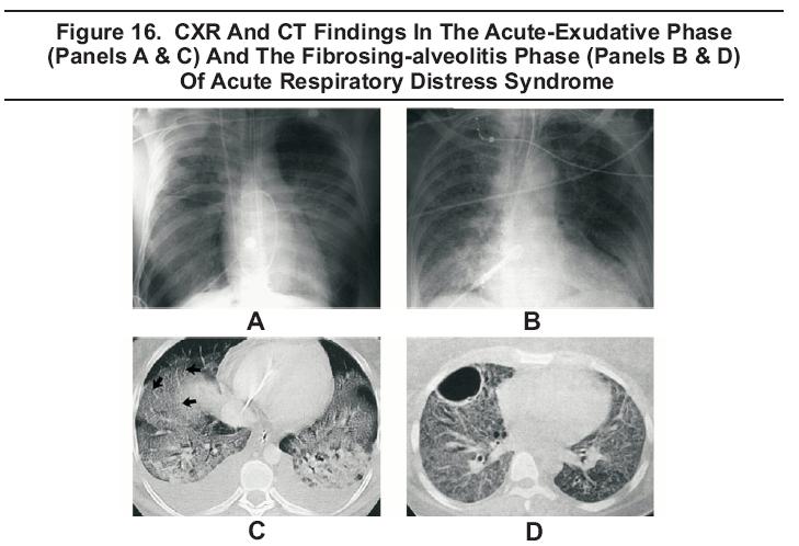
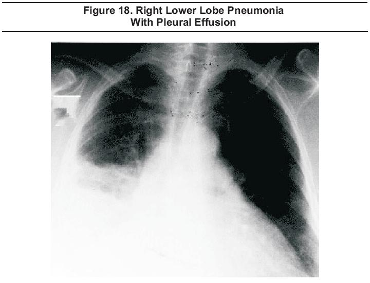
The CXR may help to determine which patients should be hospitalized. Admission is indicated when the CXR shows bilateral involvement (Figure 17), multilobar involvement, cavitation, rapid progression, or pleural effusion (Figure 18). In addition, the CXR may help in differentiating pneumonia from other conditions, may suggest specific etiologies, and may detect coexisting conditions, such as lung abscess or bronchial obstruction.

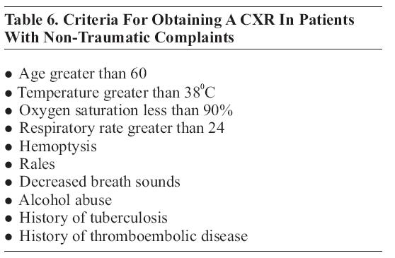
In 2002, Rothrock et al proposed a decision rule for when to obtain a CXR for non-traumatic presentations. They concluded that the presence of any of 10 criteria necessitates CXR and that, when used in this fashion, the CXR has a sensitivity of 95% and specificity of 40% for acute pulmonary pathology;11 see Table 6.
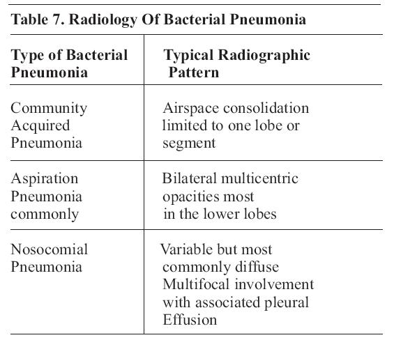
Emerman et al found that physician judgment as to when to order a CXR outperformed many decision rules with a sensitivityof 86% and a specificity of 58%.12 Traditional classification of pneumonia into lobar and bronchial is less useful than a moreclinical classification; see Table 7. 115
Chest radiography has traditionally been recommended as a part of the work up for febrile children (greater than 380C or 100.40 F) younger than three months. However, based on meta-analysis of three studies including a total of 617 infants, the chance of a positive chest radiograph in a febrile infant younger than three months of age with no pulmonary signs or symptoms was found to be only approximately 1%.10,13,14 For children older than three months of age, CXR should be considered for febrile children with temperature greater than 390 C or 102.2 0F and a WBC count greater than 20,000/mm . 3 15 CXR is usually not indicated in febrile children older than three months of age with temperature less than 390 C without clinical evidence of acute pulmonary disease.10
Chest radiograph cannot, by itself, be used to differentiate between viral and bacterial disease.116 Alveolar (lobar) infiltrate is an insensitive but reasonably specific indication of bacterial infection.116
ACR's Appropriate Criteria gives chest radiograph a highly appropriate (9/9) rating for the evaluation of HIV positive patients with cough, dyspnea, chest pain, or fever. CT of the chest is also appropriate (8/9) in HIV positive patients with acute respiratory symptoms and negative or non-specific CXR findings. If there is a high clinical suspicion for a pulmonary infection in the setting of a normal chest radiograph, a high-resolution, non-contrast CT scan may be warranted to assess for subtle abnormalities. Patients who have a normal chest radiograph and PCP will usually exhibit focal areas of ground-glass opacity on CT.5
CT is only moderately appropriate (4/9) when CXR shows diffuse infiltrates. It is highly appropriate (8/9) when non-infectious diseases are suspected. CT findings can frequently suggest the diagnosis, or at least limit the diagnostic possibilities, and may identify optimal sites for obtaining a biopsy.5
The radiological presentation of primary tuberculosis is variable. Parenchymal infiltrates anywhere in the lung fields are possible and pleural effusion may be associated or may occur alone. Hilar adenopathy is sometimes the only finding.117
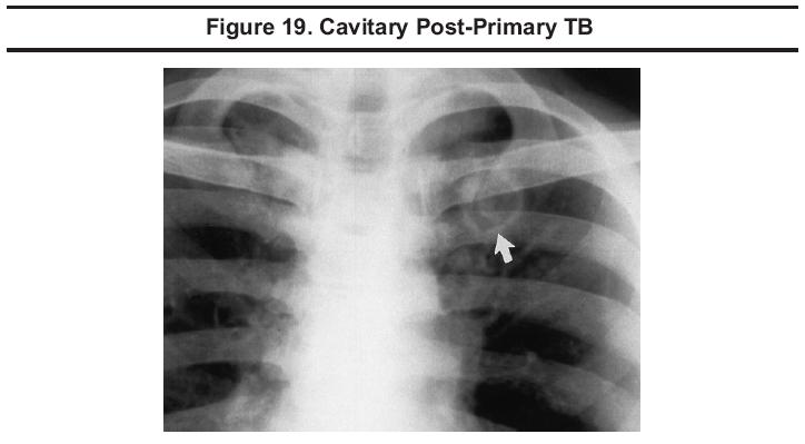
The typical radiological presentation of post-primary tuberculosis in adults (reported in 58% of patients) is with infiltration nodules in the upper zones, with or without cavitation (Figure 19).117 Pediatric post-primary pulmonary tuberculosis typically shows consolidation, cavitation, multi-focal ill-defined airspace opacities in the upper lobes, apical pleural thickening, and evidence of prior pulmonary tuberculosis.118

Screening chest radiography is indicated for patients with positive screening tests but need not include lateral views. Meyer et al compared PA CXR with PA and lateral CXR in 535 cases and found that the lateral view identified findings not present on the PA view in only 0.4% cases, and in no case did the unique findings on the lateral view alter patient management.119 High resolution CT has been used to predict activity.120
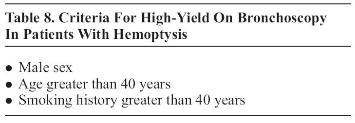
Chest radiography should be included in the initial evaluation of patients presenting with hemoptysis.121 Based on a retrospective review of records of 119 bronchoscopies performed for hemoptysis, O'Neil et al concluded that patients with less than two risk factors for malignancy and negative CXR may be managed with observation; see Table 8.121CT and bronchoscopy are complementary examinations in patients with two or more risk factors for malignancy or with persistent/recurrent hemoptysis and negative CXR.122 ACR's Appropriateness Criteria for CT in this setting is high at 8/9.6 In patients with two or more risk factors for malignancy and positive chest radiograph, ACR's Appropriateness Criteria for CT is also 8/9.6
CT may be better than bronchoscopy at defining the cause of hemoptysis; the two modalities are equally effective at determining the site of bleeding.123
Massive hemoptysis (greater than 300 mL/24 hrs) can be effectively treated with either surgery or percutaneous bronchial artery embolization.124 Embolization for massive hemoptysis receives an ACR appropriateness rating of 8/9.6
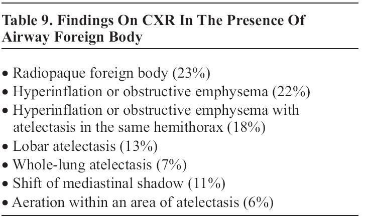
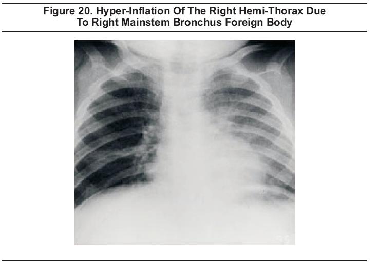
Chest radiograph findings in the presence of an airway foreign body (Figure 20) based on a retrospective chart review of 133 children are shown in Table 9.125

Non-contrast CT is easy, fast, and 100% sensitive for upper esophageal foreign bodies.126It should be the first choice for diagnostic imaging of suspected upper esophageal foreign bodies not expected to be visible on plain radiographs. CT may also detect foreign bodies not seen with barium studies.126
Echocardiography (ECHO) may be used to evaluate for wall motion abnormalities and is frequently included as a part of an emergency cardiology consultation. ECHO may also be used as an adjunct to exercise or pharmacological stress testing. The American College of Cardiology, the American Heart Association, and the American Society of Echocardiography have issued joint guidelines for clinical use.40
While there are a wide array of class I and IIa indications for adult and pediatric cardiology patients, those most pertinent for emergency department patients have to do with the evaluation of chest pain patients and establishing the diagnosis and prognosis of chronic ischemic heart disease. Per the ACC/AHA/ASE guidelines, ECHO is a class I recommendation for the evaluation of chest pain in patients with suspected acute myocardial ischemia when baseline ECG and laboratory markers are non-diagnostic and when the study can be obtained during pain or within minutes after its abatement. The use of ECHO in this way, however, generally requires the physical presence of a cardiologist trained in the use of ECHO for the detection of wall motion abnormalities early in the course of the event, which is not achievable in many centers. ECHO is not indicated for chest pain of apparent non-cardiac etiology nor is ECHO indicated for patients who have electrocardiographic changes diagnostic of myocardial ischemia/infarction.40
ECHO is also indicated for patients with chest pain who have hemodynamic instability or who are suspected of having valvular, pericardial, or aortic disease.40
Exercise or pharmacological stress ECHO is recommended as a class I intervention for the diagnosis of myocardial ischemia in symptomatic individuals or for selected patients in whom the ECG assessment is less reliable. These patients include those with intermediate pretest likelihood of coronary artery disease and digoxin use, left ventricular hypertrophy (LVH) of 1 mm or more of ST depression at rest, pre-excitation syndrome, or complete left bundle branch block (LBBB). Exercise echocardiography is also recommended at the class IIa level for the detection of myocardial ischemia in women with a low or intermediate pretest likelihood of coronary artery disease. ECHO is not indicated for the screening of asymptomatic patients with a low likelihood of coronary artery disease or as routine periodic reassessment for stable patients. Stress ECHO should not be used as a routine substitution for treadmill exercise testing in patients for whom ECG analysis is expected to suffice.40
A meta-analysis based on three studies of rest echocardiography used to assess for acute cardiac ischemia in the ED setting reported that sensitivity was excellent at 93% and specificity was rated as good at 66%.127 A subsequent report in 2002 cited the sensitivity at 91%, but noted that false negative studies may be associated with small MI's.128
Cardiac Arrest: In the setting of cardiac arrest, echocardiography can be used for the detection of cardiac motion. Patients without cardiac activity do not survive regardless of electrical activity.129,130 Echocardiography may also be used to evaluate for tamponade as a cause for pulseless electrical activity.130 Pericardial tamponade and electromechanical dissociation are truly emergent and potentially lethal cardiovascular conditions requiring emergency diagnosis.99
Nuclear Medicine Scanning:Both thallium-201 and technetium-99m radiopharmaceuticals are commonly used for myocardial scanning with imaging using single photon emission computed tomography (SPECT). Several technetium-99m labeled agents are available but the most used and studied is sestamibi.131
The ACC/AHA/ASNC Guidelines for clinical use of cardiac radionuclide imaging recommend cardiac stress myocardial perfusion SPECT at the class I level in order to identify the extent, severity, and location of ischemia in patients who are able to exercise and who have a baseline ECG abnormality that interferes with the interpretation of exercise-induced ST-segment changes, i.e., pre-excitation syndromes, LVH, digoxin therapy, or greater than 1 mm ST depression. Adenosine or dipyridamole myocardial perfusion SPECT is recommended in patients with left bundle branch block or electronically-paced ventricular rhythms and for those patients unable to exercise. The use of exercise myocardial perfusion SPECT as the initial test for patients who are considered to be at high risk (e.g., diabetics) is given a class IIa recommendation.41
The diagnostic accuracy of rest myocardial perfusion imaging in patients who have acute chest pain and normal or non-diagnostic ECGs has been found to be high. Based on eight studies between 1979 and 2002, sensitivity was reported to be 90 to 100%, specificity 60 to 92%, and negative predictive value 99 to 100%.132Studies performed in lower-risk patients have demonstrated that emergency department perfusion imaging does offer incremental value. In a large, prospective, randomized, controlled study, 2475 patients were randomized to routine care or to routine care with myocardial perfusion imaging. A significantly lower admission rate was achieved in the imaging group without reducing appropriate admission for patients with acute ischemia.133Nevertheless, there are several problems that limit the use of myocardial perfusion imaging. First, there are some technical problems in obtaining high quality images that allow differentiation of diaphragm and breast shadows from pathologic perfusion defects. Second, detection of new perfusion defects is complicated in patients with previous myocardial infarction. In many centers, availability of the test is limited to certain hours of the day, and the lack of availability of experienced readers may limit the usefulness of the scans. Finally, there continues to be some controversy over whether the injection of radioactive tracers must be performed while the patient is experiencing ischemic symptoms or whether the injection may occur soon after symptom resolution.134
Exercise SPECT myocardial perfusion imaging is preferred in patients with greater than 1 mm ST depression or pre-excitation syndrome on their resting ECG, as well as for those who have undergone percutaneous transluminal coronary angioplasty (PTCA) or coronary artery bypass grafting (CABG).131 In addition, patients with less than 1 mm ST depression on their resting ECG, those on digoxin, and those with LVH, LBBB, and ventricular pacing may be considered for SPECT myocardial perfusion imaging.131
Echocardiography performed by emergency physicians has been shown to be a reliable technique for evaluating suspected pericardial effusions (Figure 5). In a study of 515 high-risk patients with images captured on video and subsequently reviewed by cardiologists, emergency physicians detected pericardial effusion with a sensitivity of 96% and a specificity of 98%.136ECHO has been suggested for use in further evaluating ED patients presenting with a complaint of dyspnea but for whom no cause has been found after standard ED evaluation. In a prospective observational study of 103 patients who presented with new-onset dyspnea but lacked any pulmonary, infectious, hematological, traumatic, psychiatric, cardiovascular, or neuromuscular explanation for their dyspnea after ED evaluation, Blaivas found a 14% incidence of pericardial effusion and recommended the use of bedside ultrasound in the evaluation of such patients.137
Pericardial effusion appears as a dark band between the pericardium and myocardium on sub-costal view (Figure 5). Evaluation for tamponade is accomplished by looking for "swinging heart sign," right ventricular collapse during mid-late diastole, or inferior vena cava plethora.
Echocardiographic guidance for drainage of pericardial fluid is the standard of care and has a 97 to 100% success rate with a complication rate of 5% and a major complication rate of 1.2%; the most commonly reported complications are hemothorax and infection.100,138 The use of a pericardial catheter for extended drainage has become more common, with a concomitant reduction in the rate of recurrence and subsequent pericardial surgery.138
In patients suspected of having infective endocarditis, the diagnosis can be facilitated by the identification of vegetations on heart valves. Echocardiography has therefore assumed a vital role in the diagnosis of this disorder. The Duke Criteria has improved the specificity and sensitivity of the diagnosis of infective endocarditis by assigning major and minor criteria, including echocardiographic findings of an oscillating intracardiac mass or vegetation, an annular abscess or new valvular regurgitation, or prosthetic valve partial dehiscence.40,139
While both TTE and TEE can identify valvular vegetations, TEE can identify much smaller vegetations and has significantly greater sensitivity, 44% and 94% respectively. Specificity is similar for the two techniques at 98% and 100% respectively.140 When the diagnosis of infective endocarditis is suspected, obtain prompt cardiology consultation. Transthoracic ECHO may be used as the initial screening test, followed by TEE if the results are negative or equivocal.50
Chest radiograph findings of hemomediastinum include widened mediastinum (greater than 8 cm at aortic arch in the supine position), blurred aortic knob (Figure 12), left apical pleural cap, opacified aorticopulmonary window, widened right paratracheal stripe (greater than 4 mm), and left pleural effusion. Additionally, the calcium sign, consisting of a discontinuity of the calcification within the aortic knob or a separation of the calcified intima from the outer aortic border of greater than 1 cm, suggests dissection.
Chest radiograph findings are usually abnormal in the presence of aortic dissection and CXR has a reported sensitivity of 90%. The presence of a normal aorta and mediastinum on CXR decreases the probability of dissection, but does not exclude it. The negative likelihood ratio is 0.3.141Positive findings include change in aortic silhouette, widening (especially if progressive compared to old films), indistinct contour or blurred aortic knob, irregularity and separation of outer vessel wall, and intimal calcium (Figure 21). In addition, displacement of adjacent structures may be seen as the esophagus with nasogastric tube to the right and posterior, trachea to the right and anterior, or left mainstem bronchus inferiorly.141

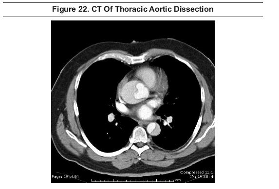
The contrast-enhanced multi-slice CT scan has become a standard test for aortic dissection.21 In fact, multi-slice CT scanning now appears to be the modality of choice for complete examination of the entire aorta.22 Computerized tomography provides excellent visualization of the aorta and branch vessels and their relationship to surrounding structures (Figure 22).21,22 Contrast is required to optimally depict the vessel lumen.51
The sensitivities of TEE, CT, and MRI for detecting dissection are similar at about 98%. TEE may provide more information on detailed anatomy of the valves in the setting of proximal dissection, and can provide functional data on regurgitation that CT cannot. By contrast, the sensitivity for TTE is only 59%.51 Specificities are 77% for TEE, 83% for TTE, 87% for CT, and 98% for MRI. CT was reported to be less effective in detecting an entry site or aortic regurgitation. Based on these findings, a noninvasive diagnostic strategy of using MRI in hemodynamically stable patients and TEE in unstable patients has been proposed.51
MRI provides at least equivalent visualization to CT and can be effectively used with or without contrast. Drawbacks include the lack of availability or poor accessibility in the emergency situation and difficulty in visualizing distal branch vessels. However, the use of MRI may obviate the need for conventional angiography in some cases.51,52
Chest radiography usually shows widening of the mediastinum, enlargement of the aortic knob, tortuosity, calcification, or tracheal deviation when there is aneurysmal dilatation of the thoracic aorta, but actual size is difficult to assess (Figure 12).142,143 However, the chest radiograph can be completely normal.144If the chest film shows abnormalities consistent with thoracic aortic aneurysm, one should have a low threshold for ordering a contrast-enhanced CT scan to better define the aortic anatomy.142,143
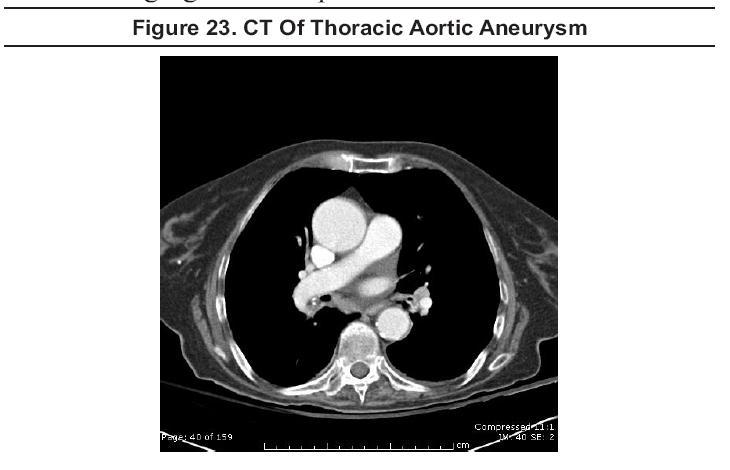
Contrast-enhanced CT scanning (Figure 23) and magnetic resonance angiography (MRA) are the preferred modalities to define aortic (and branch vessel) anatomy, and both accurately detect and size thoracic aortic aneurysms. When aneurysms involve the aortic root, MRA is preferable since CT images the root less well and is less accurate in sizing its diameter.142,143
Transthoracic echocardiography is effective for imaging the aortic root, but it does not consistently visualize the mid or distal ascending aorta or the descending aorta. TTE should generally not be used for diagnosing or sizing thoracic aortic aneurysms. TEE can visualize the entire thoracic aorta well, but, due to its semi-invasive nature, it is generally not favored for routine imaging of stable patients.143
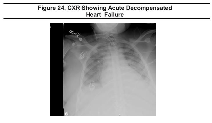
The chest radiograph (Figure 24) is rated as highly appropriate (9/9) when new onset heart failure (HF) is suspected based on symptoms and physical examination, and is rated as highly appropriate (9/9) with previously diagnosed HF and new or worsening symptoms. CXR is less appropriate (4/9) for patients with previously diagnosed HF and stable symptoms.8

Based on the analysis of the Acute Heart Failure National Registry (ADHERE) database, Collins et al reported that nearly 20% of patients admitted to the ED with acutely decompensated heart failure (ADHF) showed no signs of pulmonary congestion on chest radiography and suggested that clinicians not rule out heart failure in patients with no radiographic signs of congestion.144 While the initial ED CXR may be insensitive in predicting a hospital discharge diagnosis of ADHF, CXR is a simple test that remains helpful in the diagnosis of the majority of patients with ADHF and in establishing alternative diagnoses in many others.145
Congestive heart failure is readily diagnosed on CT obtained for other indications, but symptoms of congestive heart failure do not, of themselves, provide a sufficient indication for CT scanning (ACR appropriateness rating: 2/9). 8
Some issues in the area of thoracic imaging remain controversial and some approaches are in the process of change due to recently published research. Still others are currently undergoing intensive investigation. In this section, we will attempt to describe some of these current issues and controversies.
Ultrasound has been shown to be more sensitive than supine AP chest radiograph for the detection of traumatic pneumothoraces69and ultrasound is useful for the detection of hemothorax. In some centers, thoracic ultrasound is performed routinely along with the traditional FAST scan, creating the extended focused abdominal sonogram for trauma (EFAST).70
Ultrasonography has been reported to have greater sensitivity in detecting chest wall fractures than either clinical acumen or radiography; 80% vs 26% vs 24%.76In addition to rib fractures, this includes sternal fractures. While not commonly used for this purpose in the United States, ultrasound is a rapid and reliable method for identifying bony disruptions, especially in the superficial, readily accessible ribs and sternum.77 This application represents an opportunity for additional study by emergency ultrasound researchers.
In the past, the literature has stressed the importanceof rib fractures, especially those of the first andsecond ribs, as predictors of aortic injury. However, several studies have demonstrated no increased likelihood of aortic injury with upper rib fractures.2,75No additional imaging studies are mandated by thesefindings alone.
The clinical significance of myocardial contusion following blunt chest trauma is unknown. A number of diagnostic approaches have been used for diagnosis, including electrocardiography, serial enzyme measurement, and both TTE and TEE. TTE has proven inadequate, but TEE appears to be safe and to provide excellent quality images. Based on a retrospective study of 81 patients who received TEE in the evaluation of blunt chest trauma, Weiss et al found myocardial contusions, diagnosed by wall motion abnormalities, in almost a quarter of these patients. They noted an increase in mortality rate associated with this diagnosis.95However, a more recent prospective study by Lindstaedt et al of 180 patients with blunt chest trauma found only a 12% incidence of myocardial contusion, and none of their patients experienced any cardiac complications. They concluded that myocardial contusion is a frequent finding in polytraumatized patients, but that the outcome and prognosis is favorable.96
Since first generation scanners missed approximately a third of pulmonary emboli in one study, they could not be used alone to diagnose or exclude pulmonary embolism.27Additional tests that have been used in conjunction with early generation CT scanners included serial venous ultrasonography of the legs30 and CT venography of the pelvis and legs.31,109 With advanced generation scanners, it now appears feasible to use clinical risk stratification, D-dimer measurement, and multi-detector CT scanning to reliably and safely diagnose or exclude clinically significant pulmonary emboli.34-37A systematic review published in 2005 of 15 studies published between 1990 and 2004 containing 3500 patients found that the use of CT ruled out pulmonary embolism. An overall negative predictive value of 99.1% for a chest CT negative for pulmonary embolism was found, even though all generations of scanners were included in the review. This is a similar negative predictive value as that for conventional pulmonary arteriography. Furthermore, the use of advanced generation scanners should improve the negative predictive value.32
The chest radiograph is often included in the work-up of the hypertensive patient, presumably to evaluate for the presence of LVH. However, the CXR is insensitive for the detection of LVH and is not clearly indicated in uncomplicated cases.7 CXR is possibly indicated in patients with moderate to severe hypertension and probably should be reserved for patients with cardiorespiratory symptoms or signs.7,146 Echocardiography is the non-invasive modality of choice for the detection of the cardiac effects of systemic hypertension, the most common cause of LVH. 147,40
The Role Of CT In The Evaluation Of Pulmonary Infection In Immunocompromised Patients
If there is a high clinical suspicion for a pulmonary infection in the setting of a normal chest radiograph, a high-resolution, non-contrast CT scan may be warranted to assess for subtle abnormalities. Patients who have a normal chest radiograph and PCP will usually exhibit focal areas of ground-glass opacity on CT.5x
Electron beam computed tomography (EBCT) has been in use for many years as a means of measuring coronary artery calcium and estimating the overall coronary atherosclerotic plaque burden. EBCT has proven useful in identifying individuals with or at risk for coronary heart disease. However, there is still controversy as to the prognostic significance of calcium, as some investigators believe that the presence of coronary calcification may stabilize the atherosclerotic plaque.135
High-resolution images obtained rapidly by MDCT have recently improved image quality to the point where it may soon be possible to consider non-invasive coronary angiography as a routine clinical tool.48MDCT shows promise as a means of excluding coronary artery stenosis in a non-invasive fashion (Figure 25). Reports of the use of 16-slice MDCT for non-invasive coronary angiography have been appearing in the literature since 2002, and in 2005, reports of the use of 64-slice MDCT began to appear.
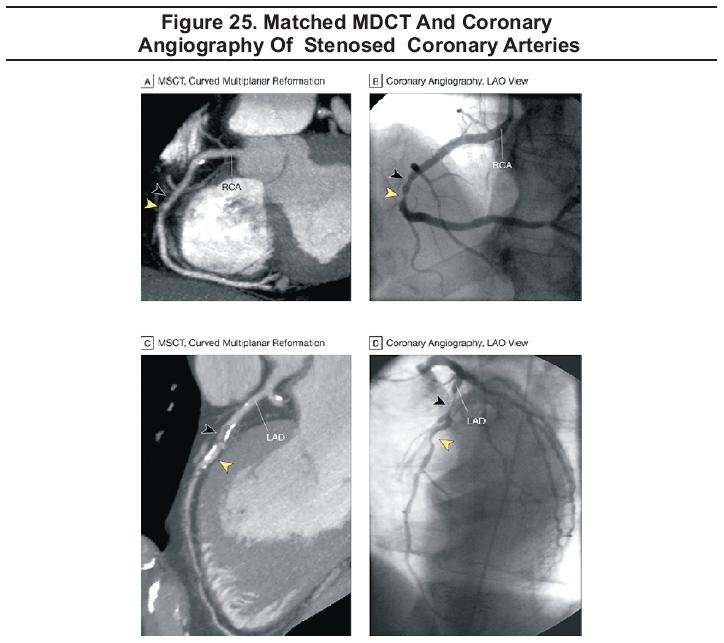
From six published or reported studies of the use of MDCT for non-invasive coronary angiography, sensitivity is reported to be 83 to 100%, specificity is 86 to 98%, positive predictive value is 79 to 87%, and negative predictive value is 97 to 100%.42-48 However, Garcia et al have recently reported the evaluation of 1629 nonstented segments in which they used 16-slice MDCT for the assessment of coronary artery stenosis. They found only 71% of the segments evaluable by MDCT. The sensitivity for detecting stenosis greater than 50% was 89% and the sensitivity for detecting stenosis greater than 70% was 94%. Negative predictive value was 99% for both categories of stenosis. Based on these findings, routine implementation of MDCT in clinical practice is not recommended, but MDCT may be useful in excluding coronary disease in selected patients in whom false positive or inconclusive stress test result is suspected.49
During each patient's ED evaluation, the emergency physician decides what, if any, imaging studies are required. For those patients who receive imaging studies, an accurate interpretation is necessary to guide treatment and disposition. Depending on the institution, imaging modality, and even time of day, studies may be read initially by the emergency physician only, by a radiology attending or resident, or by a teleradiologist. This initial interpretation is often a preliminary interpretation and definitive final interpretations are often rendered by an attending radiologist after the patient has been treated and discharged from the emergency department. There is a potential for variance between the preliminary and final interpretations.
Where there is a discrepancy between the preliminary and final interpretations, a reliable system for notification of the patient or appropriate physician is imperative. This system should minimize the medical consequences and therefore the medico-legal risk associated with an inaccurate preliminary interpretation. Routinely inform patients regarding the potential for revision of a preliminary radiological interpretation and assure reliable contact information.
In those cases where an incidental finding of potential significance is noted, such as a pulmonary nodule, notification of or referral to a primary care physician for follow up is needed. When the discrepancy is significant and would alter patient care, expeditious intervention is required. For admitted patients, physicians caring for the patient in-house should be promptly notified of the change. If the patient was discharged from the emergency department, the patient should be notified and advised as circumstances dictate. Based on the specific findings, some patients will be directed to collect a prescription while others should be advised to return to the emergency department or to contact an appropriate physician. It is essential, therefore, that a current phone number is recorded when emergency department patients are registered. Meticulous documentation of all actions and communications can mitigate medicolegal risk.
1. Making decisions based on inadequate studies.
2. Measuring the thoracic width incorrectly.
3. Not looking closely enough for pneumothorax.
4. Waiting for unnecessary films before making clinical decisions.
5. Using chest x-rays to decide whether a patient's pneumonia needs antibiotics.
6. Getting an x-ray for known asthmatics with typical exacerbations.
7. Not getting an x-ray for COPD exacerbations.
8. Looking for ventricular hypertrophy on chest x-ray in uncomplicated hypertension.
9. Ordering an ECHO inappropriately.
10. Using chest radiography to rule out dissection.
...The 62-year-old male who complained of severe tearing inter-scapular pain was of great concern to you. While intravenous labetalol and morphine were titrated, you placed a call to CT scan to expedite his imaging. Cardiothoracic surgery and the ICU were already on board when the chest CT with contrast confirmed a descending aortic dissection.
... The 28-year-old female who was post cesarean section complaining of pleuritic chest pain was also worrisome. Anticipating anticoagulation after chest CT, you concluded the examination with a rectal examination which was negative for occult blood. After confirmation of pulmonary arterial filling defects, she was anticoagulated and admitted.
... The 32-year-old male asthmatic felt better after treatment. Lung auscultation and peak flow readings were reassuring. The order for a chest radiograph placed by your nurse was cancelled and the patient was discharged.
... The 44-year-old female with dyspnea had a history of congestive heart failure but her breath sounds were diminished on the right. You ordered intravenous analgesia and obtained a portable AP chest radiograph. Review of the frontal chest film confirmed your suspicion for pneumothorax. A repeat chest radiograph after tube thoracostomy demonstrated right lung expansion; the dyspnea improved and admission was arranged.


Evidence-based medicine requires a critical appraisal of the literature based upon study methodology and number of subjects. Not all references are equally robust. The findings of a large, prospective, randomized, and blinded trial should carry more weight than a case report.
To help the reader judge the strength of each reference, pertinent information about the study, such as the type of study and the number of patients in the study, will be included in bold type following the reference, where available.
Gary R Strange; Bruce MacKenzie
November 1, 2006
Emergency Medicine Practice • CONTINUE READING
Access every issue, our complete clinical pathway library, and earn up to 190 CME credits with an annual subscription.
Stay current with a new Emergency Medicine Practice issue every month, plus unlimited access to our complete issue library, all Interactive Clinical Pathways, and up to 190 CME credits.
Accredited By


Our Partners


 678-366-7933