Headache is the fourth most common reason for emergency department encounters, accounting for 3% of all visits in the United States. Though troublesome, 90% are relatively benign primary headaches --migraine, tension, and cluster headaches. The other 10% are secondary headaches, caused by separate underlying processes, with vascular, infectious, or traumatic etiologies, and they are potentially life-threatening. This issue details the important pathophysiologic features of the most common types of life-threatening headaches, the key historical and physical examination information emergency clinicians must obtain, the red flags that cannot be missed, and the current evidence for best-practice testing, imaging, treatment, and disposition.
A previously healthy 30-year-old man presents to the ED complaining of the “worst headache of my life.” He describes it as sharp, nonradiating, with an abrupt onset 5 hours ago. You are concerned for subarachnoid hemorrhage. You provide pain medication and obtain a noncontrast CT scan of the head, which is negative. The patient is feeling better and wants to go home. You wonder whether a negative CT is sufficient to rule out an SAH or whether a lumbar puncture should be done...
A 55-year-old man with history of nonsmall cell lung cancer who is on cisplatin presents with an acute headache and lethargy for 6 hours. His vital signs are remarkable for a blood pressure of 210/120 mm Hg, heart rate of 70 beats/min, and a temperature of 36.7°C (98°F). His physical exam reveals a lethargic patient with no localizing neurologic signs and no meningismus. You order a noncontrast CT of the head and consider lowering this patient’s blood pressure, though you wonder how much and how fast it should be reduced...
A 45-year-old woman presents to the ED complaining of a severe occipital headache, neck pain, and dizziness. Earlier in the day, she was involved in a motor vehicle crash and suffered “whiplash.” Her neurologic exam is normal, including no nystagmus and normal cerebellar function, but you are concerned that this patient may have a vertebral artery dissection, and you order a CTA head and neck. You wonder: if it’s positive, should the treatment include anticoagulation or antiplatelet therapy...or both?
The third edition of the International Classification of Headache Disorders (ICHD-3), published in January 2018, is the most up-to-date and widely accepted standard criteria for the classification of headaches.1 The ICHD-3 classifies headaches into 3 distinct categories: (1) primary headache disorders, including migraine, tension, and cluster headaches; (2) secondary headaches, including potentially life-threatening forms of headaches such as those secondary to vascular disorders, traumatic injury, and disorders in hemostasis; and (3) cranial neuropathies, such as trigeminal neuralgia.
The National Hospital Ambulatory Medical Care Survey reviewed over 10,000 patients presenting to emergency departments (EDs) for acute headache and found that 2% represented secondary headaches.2 Although they are rare, life-threatening headaches require prompt diagnosis and treatment, as delays in some diagnoses can have a mortality rate approaching 50%.3
Evaluating complaints of acute headache is a common practice in the ED, and distinguishing benign from serious pathology can be a diagnostic challenge. A focused workup begins with a careful, well-organized clinical history and physical examination. Physical examination findings such as abnormal vital signs, papilledema, cranial nerve palsies, and neck pain are suggestive of more concerning headache etiologies.4 Resources such as ocular ultrasound, neuroimaging, and lumbar puncture are important strategies, but the sensitivity and specificity of the results must be understood in order to apply them correctly. This issue of Emergency Medicine Practice focuses on the most commonly encountered causes of life-threatening secondary headaches and provides best-practice recommendations on their initial evaluation and management.
A literature search from 1993 to 2018 was conducted using PubMed and Ovid MEDLINE®, with the search terms headaches AND emergency, sudden onset, fever, visual symptoms, neurologic deficits, high-risk, trauma, immunocompromised, pregnancy, coagulopathy, and life threatening. The National Guideline Clearinghouse and the Cochrane Database of Systematic Reviews were searched. Guidelines published by the American College of Emergency Physicians (ACEP) and the American Academy of Neurology were searched. International guidelines, including the Canadian and European neurology guidelines, were also reviewed. Over 500 abstracts published within the last 25 years were examined, and 89 of these full-text articles were reviewed and included for reference. Many of the identified articles were prospective studies, meta-analyses, clinical guidelines, and literature reviews.
Brain parenchyma has no pain receptors. Headaches result from traction to or irritation of the meninges and blood vessels, which are the only innervated central nervous system (CNS) structures. Nociceptors located in surrounding tissue and vasculature may be stimulated by trauma, neurogenic inflammation, edema, tension, or space-occupying lesions. It is the activation of these specific nerve ganglion complexes by neuropeptides, including substance P and calcitonin gene-related peptide, that contributes to headache pain.5
Both primary and secondary headaches have common pain pathways, so response to pain medications does not exclude potential life-threatening secondary causes. Multiple case reports document relief with nonsteroidal anti-inflammatory drugs (NSAIDs), triptans, and neuroleptics in patients with subarachnoid hemorrhage or mass lesions.6 Therefore, in accordance with the guidelines from ACEP, excluding dangerous secondary causes of headaches should not be based on response to analgesics (ACEP Level C recommendation).7
Elderly patients presenting with headache are at higher risk for secondary causes of headache, such as intracranial hemorrhage, acute angle closure glaucoma, giant cell arteritis, and malignancy.8 A large retrospective study that evaluated risk factors for intracranial pathology in patients presenting with headache found that patients aged > 50 years were 4 times more likely to have a pathologic diagnosis.4 Table 1 lists causes of secondary headaches and their initial signs and symptoms.


The following sections summarize the etiology and clinical features of the most common life-threatening headaches that should be on the differential for a patient with a headache: subarachnoid hemorrhage (SAH); cervical artery dissection (CAD), which includes vertebral artery dissection and carotid artery dissection; cerebral venous thrombosis (CVT); idiopathic intracranial hypertension (IIH); giant cell arteritis (GCA); and posterior reversible encephalopathy syndrome (PRES). Also on the differential and discussed in following sections are meningitis, acute angle closure glaucoma, pre-eclampsia/eclampsia, and carbon monoxide poisoning.
SAH represents approximately 1% of all headaches presenting to the ED and affects nearly 30,000 North Americans each year.9 Nontraumatic or spontaneous SAH is most commonly caused by aneurysm rupture; other etiologies include arteriovenous malformations and idiopathic etiologies.10 Misdiagnosis can be catastrophic, with case-fatality rates up to 50%.11
Perhaps the most distinctive historical feature of SAH is a headache described as “abrupt in onset,” a feature that is documented in 75% of SAH patients. Approximately 25% of these patients describe a transient alteration or complete loss of consciousness.12 Additional symptoms include neck stiffness, vomiting, and double vision. Approximately 20% of patients with SAH have warning signs of a sentinel bleed preceding the major hemorrhage. Symptoms often occur within days to weeks of rupture and include: (1) headaches lasting several hours or days, (2) associated cranial nerve palsies, (3) neck pain, and (4) nausea and vomiting.13,14 A recent meta-analysis of 22 studies offered important historical and examination findings seen in SAH.15 (See Table 2.)

CAD is a diagnosis that includes carotid artery dissections and vertebral artery dissections, and it is estimated to be the cause of 2% of all strokes and 20% of strokes in adults aged ≤ 50 years.16 CAD can occur spontaneously, but is more commonly associated with a traumatic event such as seat-belt injury, hanging, vigorous physical activity, and chiropractic manipulation.17 Disruption of the blood vessel wall leads to intimal bleeding, hematoma formation, and eventually thromboembolic stroke. Risk factors for CAD include Ehlers-Danlos syndrome, osteogenesis imperfecta, and Marfan syndrome.18 For more information on CAD, see the July 2016 issue of Emergency Medicine Practice, “Cervical Artery Dissection: Early Recognition and Stroke Prevention.”
CVT commonly presents as a gradual-onset headache, although some cases have been documented as CVT presenting with a thunderclap or sudden-onset headache.19 Typically the result of thrombotic disease, risk factors for CVT include oral contraceptive use, pregnancy and postpartum states, Factor V Leiden deficiency, and lupus. Infections spreading from the sinuses, ears, face, and mouth can also cause a sinus thrombus to develop.20
Typically associated with obese females of child-bearing age, the incidence of IIH has been increasing due to the rise in obesity in the population.21 Hyper-vitaminosis A from excessive dietary intake has been a long-known cause of IIH.22 Other retinoids used in the treatment of dermatologic conditions and cancer therapy, all-trans-retinoic acid, retinol, isotretinoin, etretinate, and tretinoin have also been reported to be associated with IIH. Tetracyclines have been linked to IIH in a number of cases, often appearing shortly after onset of treatment.22
GCA is a rare diagnosis with a prevalence of < 1%.23 It is 3 times more likely in women and almost exclusively found in patients older than 50 years.24 Common clinical features of GCA include systemic symptoms such as fever, fatigue, and myalgia, and localized symptoms such as headache, jaw claudication, and visual symptoms such as diplopia and amaurosis fugax.25 Polymyalgia rheumatica is present in more than half of all GCA patients; therefore, history should include questions related to chronic muscle pain, particularly in the shoulders and hips.26
PRES is a form of hypertensive emergency that is reversible.27 The presumed pathogenesis of PRES suggests that severe hypertension leads to cerebral autoregulatory failure, vasodilatation, interstitial extravasation of fluid, and subsequent vasogenic brain edema.28 The most common conditions associated with PRES are hypertensive encephalopathy, eclampsia, and the use of immunosuppressive agents such as cyclosporine, tacrolimus, and cisplatin.29 The clinical syndrome of PRES is typically characterized by elevated blood pressure, acute-onset headache, and altered level of consciousness. Generalized tonic-clonic seizures occur in up to 75% of these patients.30
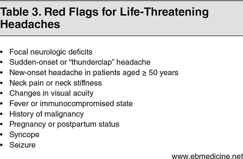
The prehospital approach to the patient with headache includes: (1) conducting a primary survey and eliciting a basic history, particularly with regard to the time of onset, provocation, quality, and severity; (2) conducting a focused neurologic examination using the Cincinnati Prehospital Stroke Scale (CPSS); and (3) assessing for red flags; ie, signs and symptoms of dangerous causes of headache. (See Table 3.)
The CPSS provides a framework for the prehospital assessment of acute headache, as several life-threatening headaches present with neurologic deficits.31 Patients with neurologic deficits and patients with severe, sudden-onset headache should be transported immediately to the nearest available stroke center. Acetaminophen may be offered for initial pain management during transport; opioids and NSAIDs should be avoided.
A thorough history and physical examination will guide the emergency clinician in determining the need for laboratory testing and imaging studies.
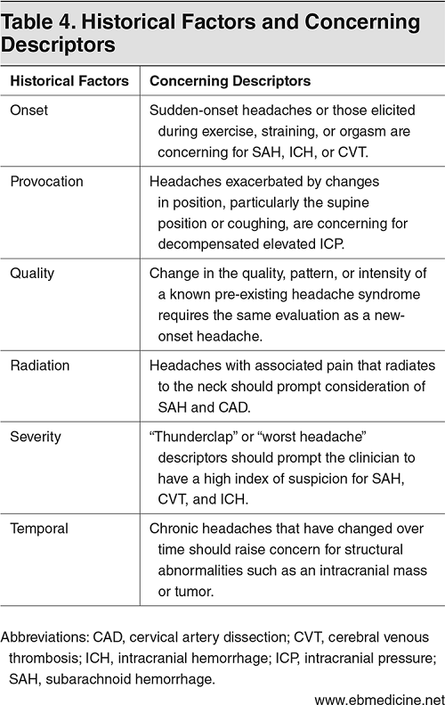
The history should include the time of onset of the headache and its location, its severity, and associated symptoms. Table 4 lists historical factors that are concerning for a life-threatening etiology. Associated signs and symptoms, particularly those considered to be red flags for life-threatening headaches, should be considered when eliciting the history of a patient presenting with headache. (See Table 2 and Table 3.)
The physical examination in the patient with headache includes an assessment of the following 5 components: (1) vital signs, (2) neurologic function, (3) cranial nerves, (4) head and neck, and (5) fundoscopic evaluation.
A complete set of vital signs must be obtained in all patients presenting to the ED with an acute headache. Abnormal vital signs, particularly with regard to elevated blood pressure and temperature, may be associated with life-threatening etiologies.
Acute headache in the setting of severe hypertension should prompt a search for signs of end-organ damage such as hypertensive encephalopathy, intracranial hemorrhage, PRES, and pre-eclampsia in pregnant women.
Patients presenting with increased intracranial pressure (ICP) may have an associated vasopressor response. This is also known as the Cushing reflex, which is a triad of increased blood pressure, irregular respirations, and bradycardia. These findings should prompt the emergency clinician to look for causes of ICP that include SAH, acute stroke, and IIH.
Fever in the setting of acute headache should raise concern for CNS infections that include meningitis, brain abscess, and encephalitis. In particular, patients presenting with headache, fever, altered mental status, and neck stiffness should prompt consideration of meningitis. Ninety-five percent of patients with bacterial meningitis will present with a combination of 2 of these 4 symptoms.32
A focal neurologic deficit in the setting of an acute headache is the single highest predictor for the presence of intracranial pathology.33 Careful attention should be paid to evaluating the patient’s mental status, including level of alertness, orientation, and attention (eg, reciting the months of the year in reverse). The neurologic examination should also include assessment of motor strength, coordination, reflexes, sensory function, and gait. Neurologic abnormalities suggestive of a lesion involving the anterior circulation (such as dysarthria and cognitive impairment) should prompt consideration of carotid artery dissection, particularly if Horner syndrome is also present. Conversely, lesions of the posterior circulation can cause dizziness, vision changes, and limb weakness and are symptoms that may be seen in strokes from a vertebral artery dissection.34
The focused neurologic examination in patients with a chief complaint of headache includes a careful assessment of select cranial nerves (CN). The optic nerve (CN II) controls the afferent pupillary reflex and can be tested using the swinging flashlight test, which involves shining a bright light into each eye and watching for brisk pupillary constriction. Paradoxical dilation indicates an afferent pupillary defect, also known as a Marcus Gunn pupil. Conditions with an afferent pupillary defect include optic neuritis, GCA, and central retinal artery occlusion.35
CN III, IV, and VI are tested by having the patient follow, with his eyes, a finger that is drawing an “H” pattern without moving the head. In the setting of headache, oculomotor nerve palsy (CN III), ipsilateral mydriasis, ptosis, and abnormal extraocular movements (“down-and-out eye”) are concerning for a posterior communicating aneurysm and can also be seen in the setting of SAH.36
Abducens nerve (CN VI) deficits may be elicited when a patient develops diplopia with lateral gaze. The patient may also have convergent strabismus, where one eye points toward the nose. CN VI deficits may be present in patients with ICP, such as with IIH and CVT. These patients may also have impaired visual acuity, visual field defects, and tunnel vision due to papilledema.37
A thorough head and neck examination should accompany the physical examination for all patients presenting with headache. Findings such as neck rigidity and stiffness should raise concern for possible meningitis or SAH.
Patients presenting with headache and neck pain should raise concern for CAD. Partial Horner syndrome may also be present in patients with CAD, leading to miosis and ptosis. Anhidrosis is absent, as sympathetic fibers innervating the facial sweat glands are not affected. Patients may present initially with head or neck pain but without any neurologic deficits, as neurologic symptoms may take several days to develop.38
Unilateral tenderness and beading to the temporal artery in a patient presenting with acute headache may suggest GCA. Smetana et al published an excellent review on clinical examination findings for GCA. Symptoms with the highest positive likelihood ratio (LR) include jaw claudication (LR, 4.2), diplopia (LR, 3.4) Physical examination findings include temporal artery beading (LR, 4.6), and temporal artery tenderness (LR, 2.6).24
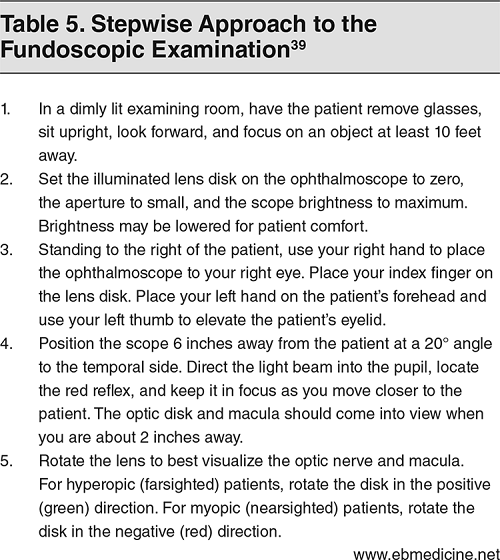
Examination of a patient presenting with acute headache should include a fundoscopic examination. (See Table 5.) If possible, pharmacologic dilation will enhance visualization of the disk, macula, and proximal vessels. Dilation can be achieved by using 1 drop of 1% tropicamide. Fundoscopy can reveal papilledema, a common finding in IIH, malignant hypertension, and CVT.
Video demostration of fundoscopic examination:
Routine laboratory testing, such as a complete blood cell count and a metabolic panel, are typically of low utility in aiding in the diagnosis of headaches. However, if a life-threatening headache is suspected, there are several circumstances that warrant specific laboratory testing. For example, there is utility in obtaining a serum blood sugar level for a patient with headache and altered mental status, or a pregnancy screen for a female patient with acute headache and elevated blood pressure.
Erythrocyte sedimentation rate (ESR) and C-reactive protein (CRP) are often obtained on patients being evaluated for GCA. However, several retrospective studies have demonstrated their poor sensitivity and specificity to diagnose GCA.40,41 While an elevated ESR and CRP makes the diagnosis of GCA more likely, patients for whom there is a high suspicion of GCA should be treated, and consultation obtained for a biopsy even if both the ESR and CRP results are negative.
If there is concern for carbon monoxide (CO) exposure, carboxyhemoglobin (COHb) level can be obtained with a co-oximeter, a device that spectrophotometrically reads the percentage of total hemoglobin saturated with CO (blood gas).42 Either a venous or arterial blood sample can be used to detect COHb.
Several small studies have looked at the utility of D-dimer to risk stratify patients presenting to the ED with headache suspicious for CVT.43 One prospective study that looked at > 300 patients determined that a D-dimer level < 500 mcg/L had a 97% sensitivity and a negative predictive value of 99%.44 A 2015 retrospective study and literature review of more than 600 patients found similar sensitivities and negative predictive value.45 Thus, D-dimer may be used to exclude CVT in low-risk patients (defined as headache patients with a normal neurological examination, normal standard head computed tomography (CT) scan, and absence of risk factors such as pregnancy or puerperium.46 High-risk patients, such as those who are pregnant or those with active malignancy, should proceed to magnetic resonance venography.
Despite the frequent utilization of brain CT, its diagnostic yield is low, ranging from 2.5% to 10% in patients presenting with nontraumatic headaches.47,48 Many disease processes will likely not be identified on CT scan, such as carbon monoxide poisoning, IIH, or meningitis/encephalitis. Noncontrast head CT should be obtained in patients suspected of having an acute intracranial hemorrhage; contrast-enhanced CT should be considered when there is concern for a vascular lesion or a space-occupying lesion (eg, tumor or abscess) or infection (eg, toxoplasmosis or neurocysticercosis).
CT angiography or magnetic resonance imaging (MRI) of the head and neck are the 2 recommended initial imaging modalities for CAD.49 Digital subtraction angiography has classically been used as the gold-standard imaging for vascular dissections; however, it is invasive, resource-intensive, and has decreased sensitivity, particularly with regard to luminal abnormalities.49
In cases of suspected CVT, a noncontrast CT may be the initial imaging obtained and may help rule in or rule out an alternative diagnosis. However, magnetic resonance venography is recommended when clinical suspicion for CVT is high.50
For suspected PRES, neuroimaging is the most important diagnostic tool. Typical findings are symmetrical white matter edema in the posterior cerebral hemispheres.51 Although vasogenic edema can be visualized in some patients using noncontrast CT, brain MRI is much more sensitive.51,52
Current ACEP Clinical Policy recommendations regarding emergent neuroimaging on patients presenting to the ED with acute headache are:7
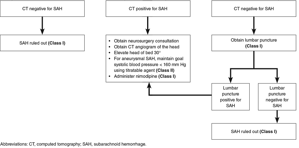
Patients with a sudden-onset, severe headache who have negative findings on a head CT, normal opening pressure, and negative findings in cerebrospinal fluid analysis do not need emergent angiography and can be discharged from the ED, with follow-up recommended. (ACEP Level B recommendation.) A recent prospective study found that the combination of a negative head CT and negative lumbar puncture has been found to have a negative predictive value of 100%.53
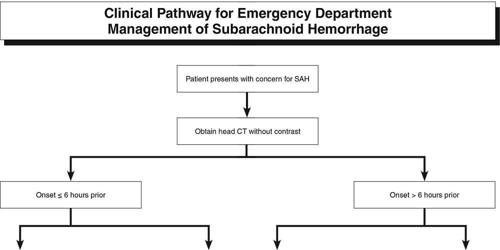
Perhaps the greatest limitation of CT for the diagnosis of SAH is that its sensitivity is time-dependent. Recent literature, including a prospective study that enrolled more than 3000 patients, found the sensitivity of CT conducted within 6 hours of symptom onset to have a sensitivity of 100% (97%-100%), a specificity of 100% (99.5%-100%), and a negative predictive value of 100% (99.5%-100%).54 Two large 2016 meta-analyses demonstrated that CTs that were conducted within 6 hours of headache onset and whose results were read by an attending radiologist as negative had a LR of 0.01, essentially ruling out the diagnosis of SAH.16,55
The 2008 ACEP Clinical Policy Guidelines (currently under revision) as well as the 2012 AHA Guidelines recommend a lumbar puncture in all patients being evaluated for SAH who have a negative noncontrast CT, regardless of time of onset (Level B recommendation).7,56 However, the most up-to-date literature supports a CT-only approach in patients presenting within 6 hours of headache onset.54,55
Lumbar puncture may be utilized as a diagnostic tool in patients with headache and historical or examination findings suggestive of SAH, IIH, or meningitis. Lumbar puncture carries the risk of possible herniation, particularly in patients with headache and signs of increased ICP (eg, papilledema, absent venous pulsations on fundoscopic examination, altered mental status, focal neurologic deficits, or signs of meningeal irritation). Patients exhibiting these symptoms should undergo a neuroimaging study before having a lumbar puncture. In the absence of clinical findings suggestive of increased ICP, a lumbar puncture may be performed without obtaining a neuroimaging study. (ACEP Level C recommendation)
Patients with suspected IIH should have an opening pressure obtained with the patient lying in the lateral decubitus position, as performing the test on a patient in the sitting position can give falsely elevated readings. Patients with IIH have normal cerebrospinal fluid composition and, often, an elevated opening pressure (> 25 cm H2O). Removal of a volume of cerebrospinal fluid can provide temporary relief of symptoms. Removal of 1 mL of cerebrospinal fluid will lower the cerebrospinal fluid pressure by approximately 1 cm H2O.57
Lumbar puncture findings for SAH include an elevated opening pressure > 20 cm H2O and a persistent number of red blood cells (RBCs), usually in the thousands, from tubes 1 through 4. There is no guideline for a cutoff number of RBCs to diagnose SAH. A 2013 retrospective study looked at over 4000 patients who received a lumbar puncture for suspicion of SAH. The study found no patients with SAH who had RBCs < 100 in the final tube; RBCs > 10,000 increased the odds of SAH by a factor of 6.58
Xanthochromia is produced by the breakdown of hemoglobin, and it produces a yellow-tinged color in the cerebrospinal fluid. Xanthochromia is time-dependent, and takes 2 to 12 hours to develop, so its absence in patients presenting within this timeframe may not be helpful. A 2015 prospective study found that patients with no xanthochromia and < 2000 RBCs were effectively ruled out of having SAH, with a combined sensitivity of 100%.59
Patients with a sudden-onset, severe headache who have negative findings on a head CT, normal opening pressure, and negative findings in cerebrospinal fluid analysis do not need emergent angiography and can be discharged from the ED, with follow-up. (ACEP Level B recommendation.)
With respect to IIH, brain imaging with CT or MRI can be helpful in excluding other causes of secondary headache that may mimic IIH, such as CVT and intracranial mass. However, the brain parenchyma and ventricles generally appear normal on CT in patients with IIH.60
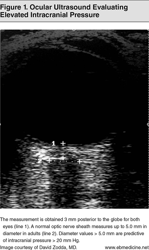
Ocular ultrasonography can expedite the diagnosis and management of a variety of ocular emergencies in the setting of headache and can also be potentially used to assess for increased ICP and thus increase the safety of performing a lumbar puncture without CT. Several studies have demonstrated good correlation between ICP and sonographic optic nerve sheath diameter measurement.61,62
A normal optic nerve sheath diameter measures up to 5.0 mm in adults. The measurement is obtained 3 mm posterior to the globe for both eyes. Three measurements should be obtained and averaged. Values > 5.0 mm are predictive of ICP > 20 mm Hg; increased ICP should be suspected with values above this threshold.63 (See Figure 1.)
There is not enough evidence at this time to completely exclude increased ICP with normal optic sheath diameters on bedside ocular ultrasonography; thus, if clinical suspicion is high for increased ICP, brain CT should be performed first before proceeding with a lumbar puncture.
Initial treatment for patients presenting with suspicion of a severe life-threatening headache includes a primary assessment of their airway, breathing, and circulation. Patients should be in a monitored setting and vital signs obtained quickly. Often, blood pressure normalizes once pain is treated. Patients with persistently elevated blood pressure and signs of neurologic end-organ damage (eg, confusion, lethargy, seizure) should be evaluated for potential hypertensive emergency. These patients need emergent blood pressure management.
The treatment goal for hypertensive emergencies is to lower the mean arterial pressure (MAP) = one-third systolic blood pressure + two-thirds diastolic blood pressure by approximately 25% in the first hour. If acute ischemic stroke is suspected, the blood pressure should be lowered below 185/110 mm Hg prior to the administration of thrombolytic therapy (for those patients who are candidates for thrombolytic therapy).64
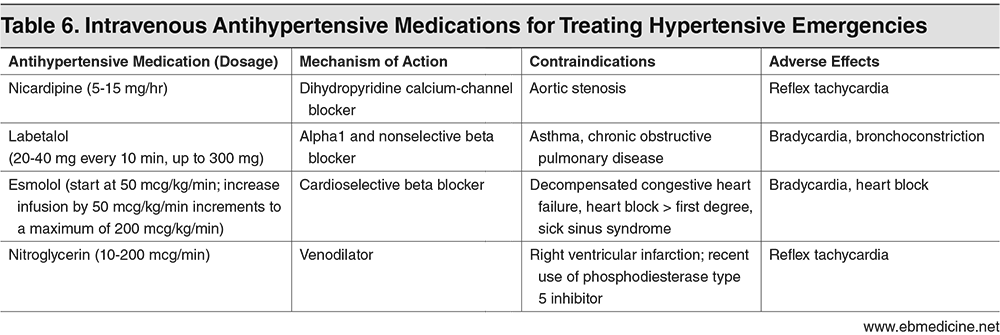
Results from the INTERACT-2 and ATACH-2 trials demonstrated that intensive blood pressure management does not appear to impact rates of death or disability. Rather, the INTERACT-2 trial demonstrated that a target systolic blood pressure (SBP) < 140 mm Hg was associated with improved functional outcomes. Based on this, guidelines recommend that for ICH patients presenting with SBP between 150 and 220 mm Hg and without contraindication to acute blood pressure treatment, acute lowering of SBP to 140 mm Hg is safe. (Class I, Level A recommendation.)65,66 The choice of antihypertensive agent and the goals of therapy should be tailored to the individual patient, with careful consideration of contraindications and adverse effects. (See Table 6.)
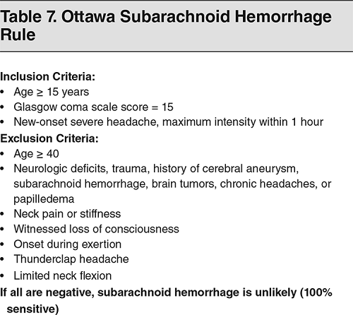
Several clinical decision tools have been developed to aid in risk stratifying patients for SAH. The most well-known tool is the Ottawa subarachnoid hemorrhage rule. (See Table 7.) The original Ottawa SAH rule study demonstrated 100% sensitivity for SAH, and 15.3% specificity.67 This study has since been prospectively validated, and although it has been challenged for its interobserver variability, it does provide helpful red flags for clinicians to consider when evaluating patients with headache.68,69
A neurosurgical consultation and CT angiogram of the head should be obtained immediately when SAH is diagnosed. ED management focuses on monitoring the patient’s airway, breathing, circulation, and mental status. Appropriate analgesia and antiemetics should be provided as needed. Elevating the head of the bed to 30° may improve venous drainage.
For aneurysmal SAH, the American Stroke Association guidelines currently recommend obtaining a target goal SBP of 160 mm Hg with a titratable agent that includes nicardipine or clevidipine.56
Nimodipine, a calcium-channel blocker, should be provided to patients with aneurysmal SAH to improve neurologic outcomes. Nimodipine is typically administered 60 mg orally every 4 hours. The use of prophylactic antiepileptic drugs is controversial, and little literature has been published recently. One study demonstrated worse outcomes based on the Glasgow Outcome Scale when anti-epileptic drugs were administered. Phenytoin accounted for about half of the antiepileptic drugs used in that study.70 Levetiracetam is used frequently, though data are limited. Anticonvulsant prophylaxis may be considered in the immediate posthemorrhagic period and should be limited to a 3- to 7-day course. Longer courses may be considered for patients with prior seizure, intracerebral hematoma, intractable hypertension, infarction, or aneurysm at the middle cerebral artery.56,71

Treating CVT focuses on managing the initiating cause of the thrombosis. Broad-spectrum antibiotics should be employed if an infectious source is suspected. Treating CVT with anticoagulation is controversial, as it is common for CVT to present with intracerebral hemorrhage or hemorrhagic transformation.19,72 Ferro et al documented that as many as one-third of these patients had hemorrhage on CT or MRI.20 However, the data may be slightly biased, as providers may have avoided anticoagulation in patients with poorer prognosis;72 more robust studies are needed. Despite the controversy and the significant risk of bleeding, per guideline recommendations, anticoagulation is the standard therapy for this disease, with full-dose anticoagulation of low-molecular-weight heparin (eg, enoxaparin) or heparin bridge to warfarin therapy.72,74
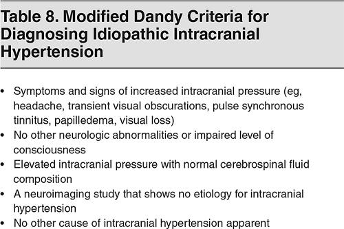
The modified Dandy criteria can aid in the clinical diagnosis of IIH.75 (See Table 8.)
A lumbar puncture can be performed for not only diagnostic purposes, but also for therapeutic purposes, although the relief is typically temporary.76 Weight loss may be recommended for obese IIH patients. A prospective study of obese IIH patients found that weight loss led to reduced symptoms, signs, and ICP.77
Acetazolamide, 250 mg to 500 mg orally twice daily, is considered the first-line pharmacotherapy for IIH. Acetazolamide is a carbonic anhydrase enzyme inhibitor that decreases cerebrospinal fluid production in the choroid plexus, thereby lowering ICP. A multicenter double-blinded randomized controlled trial found that the use of acetazolamide with a low-sodium weight-reduction diet resulted in modest improvement in visual field function.78 Potential adverse effects of the drug include flushing, hypersensitivity reactions (eg, Stevens-Johnson syndrome/toxic epidermal necrolysis or agranulocytosis). If acetazolamide therapy fails or there is a contraindication to its use, topiramate or furosemide are alternative therapies. Starting doses are not well established, but typical regimens may be topiramate 25 mg orally daily and furosemide 20 mg orally daily.79
Surgical options are available for refractory cases, such as cerebrospinal fluid shunting, venous sinus stenting, and optic sheath fenestration.
If PRES is caused by a specific medication (such chemotherapy drugs), this medication should be discontinued temporarily. The management of hypertensive episodes and maintenance of normal blood pressure is an essential component of treatment.80 The choice of antihypertensive drugs is up to the discretion of the treating clinician and includes vasodilators such as nitroglycerin, nitroprusside, or particularly nicardipine, due to its predictable response in primary neurologic events, or adrenergic inhibitors such as labetalol. Starting doses are variable and should be based on patient-specific factors including age, current blood pressure, and heart rate.29 Nitroglycerin is contraindicated when there is a concern for increased ICP. A reduction of MAP by 25% within the first hour is a reasonable goal.
Whether traumatic or spontaneous, medical management for stroke prevention is the mainstay of treatment for carotid artery dissection and vertebral artery dissection. The superiority of antiplatelets versus antithrombotics is not established. The landmark CADISP study found no difference in mortality or neurologic improvement when anticoagulation versus antiplatelet therapy were compared.81 Based on the best available evidence, we recommend using intravenous (IV) heparin followed by warfarin/direct oral anticoagulant in patients with extracranial dissections, and to use antiplatelet therapy (aspirin or clopidogrel) in patients with intracranial dissections and in patients in whom systemic anticoagulation is contraindicated.82
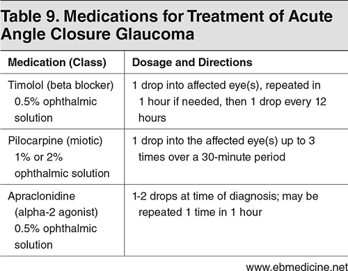
According to the American Optometric Association Consensus Panel, acute angle closure glaucoma is typically unilateral and most commonly found in either elderly or hyperoptic patients. Signs and symptoms may include headache, pain, redness, tearing, photophobia, nausea/vomiting, blurred vision, and seeing halos around lights. Pain has been noted to be associated with the rapid rise in intraocular pressure (IOP). IOP that increases above the normal usual range (10-21 mm Hg) can lead to rapid progression of symptoms, which may even include vision loss. Immediate treatment is essential, and includes consultation with an ophthalmologist. Treatment of acute angle closure glaucoma includes prompt lowering of IOP in order to preserve the patient’s vision. Options for IOP reduction include medical (pharmaceutical) therapy, laser therapy, and surgery. Medication therapies in an acute attack include topical miotics, beta blockers, and alpha agonists. Treatment options are outlined in Table 9.83-85
Patients for whom there is a high level of suspicion for GCA should be treated with high-dose methylprednisolone 15 mg/kg/day IV for 1 to 3 days, followed by oral prednisone 40 mg/day. Arrangements should be made to obtain a biopsy, even if both ESR and CRP are negative.86
The American College of Obstetricians and Gynecologists (ACOG) recommends that the diagnosis of pre-eclampsia be based on hypertension and associated symptoms and/or laboratory findings.87 (See Table 10.) Postpartum pre-eclampsia may occur up to 4 weeks post partum.88
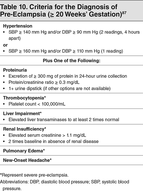
Pre-eclampsia is defined as elevated blood pressure with proteinuria or other severe symptoms in a pregnant patient at ≥ 20 weeks‘ gestation. Proteinuria is no longer necessary to diagnose pre-eclampsia if other severe symptoms are present. Severe pre-eclampsia is defined as pre-eclampsia plus 1 of the following symptoms: thrombocytopenia, liver or renal impairment, pulmonary edema, or new-onset headache.87
Treatment of severe pre-eclampsia includes IV magnesium sulfate and antihypertensive agents. Typical dosing of magnesium sulfate includes a 4- to 6-gram load given over 15 to 20 minutes, immediately followed by an infusion of 1 to 2 grams/hour.89 Patients without findings to suggest severe pre-eclampsia do not require IV magnesium, but should receive antihypertensive treatment. Agents and starting doses may include labetalol 10 to 20 mg IV, hydralazine 5 mg IV, or nifedipine 10 to 20 mg orally.87
An important aspect of medical care is to determine the acceptable threshold for testing and the potential miss of a life-threatening condition. In other words, at what threshold does further testing do more harm than good? A miss rate of 1% in many diseases is deemed acceptable. Once this threshold of 1% is crossed, further testing may cause harm to the patient rather than providing a benefit.
This leads to the need for risk stratification and the use of a shared decision-making model. Thus, a negative head CT in a patient presenting with headache within 6 hours of onset does not rule out SAH, but it does risk stratify the patient to a group with a < 1% chance of having SAH. The emergency clinician and the patient should negotiate a plan for further diagnosis and management in line with the patient’s values and preferences, with the clinician presenting the information and discussing the risks and benefits of further testing if the initial CT is negative, as well as all the risks associated with a lumbar puncture. This strategy applies not just to SAH, but to other neurologic emergencies, including meningitis/encephalitis, IIH, and hyperbaric oxygen therapy for CO poisoning.
Emergency clinicians are encouraged to obtain emergent consultation on life-threatening headaches that are diagnosed in the ED. Consultations are dependent upon the etiology of the specific medical condition and may include consultations from neurosurgery, ophthalmology, obstetrics, and infectious disease. Nearly all patients diagnosed with a severe, life-threatening headache will require admission to or transfer to a facility with access to a 24-hour neurology critical care team. These patients often require intensive care unit admission for frequent monitoring of airway, breathing, and circulation, and frequent neurologic assessment. Efforts should be made to transfer these patients to the intensive care unit as efficiently and expeditiously as possible, as delays in transfer and ED boarding have been shown to lead to increased rates of morbidity and mortality.
Headache is one of the most common chief complaints in patients presenting to the EDs in the United States. Secondary headaches represent approximately 2% of all headache presentations to the ED, and though they are rare, they can represent life-threatening emergencies. These headaches may be the result of vascular, infectious, or traumatic etiologies.
The most important factor in evaluating a patient presenting to the ED with headache is to obtain a thorough history and physical examination. Historical clues and examination findings are the foundation of the assessment and treatment plan, and should help guide decisions with regard to obtaining laboratory and imaging studies and patient disposition. Advances in diagnostic testing and imaging, such as the 100% sensitivity of CT to rule out SAH within 6 hours, can aid emergency clinicians in their workup and assessment of life-threatening headaches.
Emergency clinicians are on the front lines of cutting-edge critical care management. We now have more access to diagnostic imaging and treatment modalities than ever thought possible. And yet, perhaps the most important tools we have are our ability to listen to our patients, conduct a quality examination, and incorporate evidence-based decision-making into our clinical practice.
1. “I got a CT scan, and the lumbar puncture revealed no xanthochromia, so I discharged him, thinking he didn’t have a SAH.”
Xanthochromia is time-dependent and takes 2 to 12 hours to develop, so its absence in patients presenting within this timeframe may not be helpful. If the diagnosis for SAH is still unclear after noncontrast CT and lumbar puncture, additional diagnostic imaging may include CT angiogram and magnetic resonance angiography.
2. “When evaluating the pregnant patient for CVT, I didn’t want to subject her to any radiation, so I obtained a D-dimer in lieu of a CT scan.”
The diagnosis of CVT should be made using the clinical examination and imaging studies. Several small studies have looked at the utility of D-dimer to screen patients presenting to the ED with headache suspicious for CVT. Pregnancy is a risk factor for CVT, so this patient is not low-risk.
3. “My patient complaining of headache and neck pain had no focal neurologic deficits, so I had a very low clinical suspicion for carotid or vertebral artery dissection.”
Patients with CAD may initially present with head or neck pain, but without any neurologic deficits; the goal is to diagnose and treat before the dissection causes thrombus, which can embolize and cause stroke.
4. “The 55-year-old patient I evaluated for a new-type headache had no neurologic deficits, so I suspected that the etiology was benign, and I did not obtain imaging.”
ACEP Clinical Policy recommends that patients aged > 50 years who present with a new type of headache and a normal neurologic examination should be considered for an urgent neuroimaging study (Level C recommendation).
5. “She was 7 days post partum and came in complaining of new-onset headache and with a blood pressure of 186/92 mm Hg. Her urinalysis was negative for protein, so I ruled out pre-eclampsia.”
ACOG recommends that diagnosis of severe pre-eclampsia includes new headache and hypertension. Proteinuria is no longer necessary to diagnose pre-eclampsia if other symptoms are present. Postpartum pre-eclampsia and eclampsia may occur up to 4 weeks post partum. Treatment with IV magnesium and antihypertensives is indicated for this patient.
6. “My 60-year-old patient presented with signs of an anterior stroke; however, her last known well time was 12 hours ago, so I did not consult neurosurgery, since she was out of the window for thrombolytics.”
The 2018 AHA Guidelines for endovascular therapy in acute ischemic stroke recommends that, in select patients with signs of acute stroke whose onset is within 6 to 24 hours, mechanical thrombectomy is reasonable (Level IIa recommendation).
7. “I was evaluating a patient I highly suspected of having GCA, but both the ESR and CRP were negative, so I was able to rule it out.”
ESR and CRP are poor screening tests for GCA. While a greater ESR and CRP makes the diagnosis of GCA more likely, when there is high suspicion of GCA, it should be treated, and consultation for possible biopsy obtained, even if both ESR and CRP are negative.
8. “I have a patient I’ve diagnosed with PRES that I think is caused by her tacrolimus. I was able to manage her blood pressure, so I told her she can continue her tacrolimus.”
If PRES is caused by a specific medication, it should be discontinued temporarily. Immediate follow-up should be scheduled with the provider who prescribed the medication.
9. “I am suspecting IIH in a 27-year-old woman with headache and bilateral blurry vision. However, her vision is 20/20 bilaterally, so I was less concerned for IIH.”
Subjective blurry vision in IIH is due to papilledema, but visual acuity is typically preserved in these patients. Visual fields are affected first. If the clinician has been trained, bedside ultrasound should be attempted to assess for optic sheath enlargement.
10. “He presented to the ED with a new-onset headache, but it was mild and the neuro exam was normal. His only past medical history included HIV, so I treated him for pain and discharged him without obtaining imaging.”
The ACEP Clinical Policy on the evaluation and management of adult patients presenting to the ED with acute headache includes emergent neuroimaging on HIV-positive patients with a new type of headache (Level B recommendation).
Given the history your first patient provided, your suspicion and immediate evaluation for subarachnoid hemorrhage was appropriate. The most recent literature supports a CT-only approach in patients presenting within 6 hours. Because this patient’s symptoms had resolved, additional imaging and neurology consultation were not indicated. You utilized a shared decision-making strategy with the patient and discussed the risks and benefits of obtaining additional testing. You discharged him and cautioned him that he should return to the ED if his headache returned.
Regarding your second patient, you recognize that this cancer patient’s change in mental status and severely elevated blood pressure was likely the result of PRES. You obtained a CT of the head, which revealed white-matter changes in the posterior cerebral hemispheres. Utilizing IV nicardipine, you lowered the patient’s MAP by 25% over the first hour. In addition, you temporarily discontinued his chemotherapy medication. He subsequently became more alert and responsive.
Based upon your third patient's history of a motor vehicle crash, particularly her complaints of headache, neck pain, and dizziness following the neck trauma, you obtained a CT angiogram of the neck. The imaging revealed an extracranial dissection of the left vertebral artery. You promptly initiated anticoagulation therapy utilizing IV heparin to be followed by oral warfarin. In addition, you obtained a vascular surgery consultation and admitted the patient to the surgical intensive care unit.
Evidence-based medicine requires a critical appraisal of the literature based upon study methodology and number of subjects. Not all references are equally robust. The findings of a large, prospective, randomized, and blinded trial should carry more weight than a case report.
To help the reader judge the strength of each reference, pertinent information about the study, such as the type of study and the number of patients in the study is included in bold type following the references, where available. In addition, the most informative references cited in this paper, as determined by the author, are highlighted.
Accredited By


Our Partners







 678-366-7933


 Product Library
 Download PDF