Acute Appendicitis - Visual Diagnosis
Click to check your cart0

Visual Diagnosis

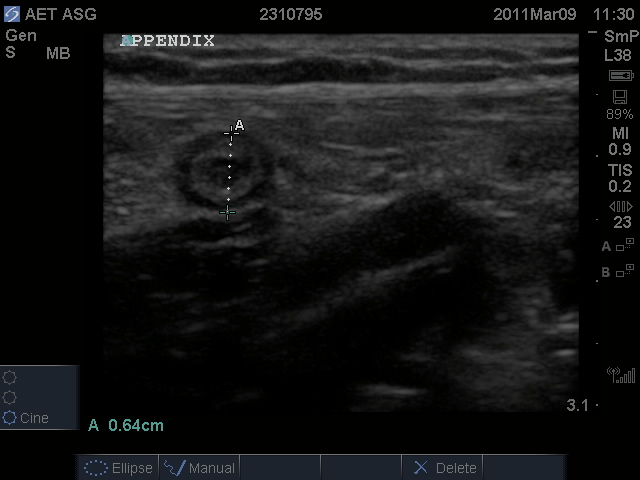
Case: A 13-year-old boy presents to the ED with right lower quadrant pain and vomiting for 1 day. Ultrasound was obtained for evaluation and is pictured here.

Acute appendicitis ultrasound in a 13-year-old-boy

Diagnosis:

This patient has acute appendicitis.

This patient has acute appendicitis based on clinical presentation and ultrasound diagnosis. Computed tomography has been the imaging modality of choice to confirm diagnosis; however, recent trends favor the use of ultrasound in children to decrease radiation exposure without risking the accuracy of the diagnosis. Once the diagnosis is confirmed, surgery consultation should be notified for further intervention.

Clinical Practice Pearls:

  • For suspected appendicitis, a linear or abdominal probe may be used on ultrasound to confirm the diagnosis.

  • Start the ultrasound scan at the point of maximum tenderness on physical examination to ensure a greater chance of detection of appendicitis.
  • Appendicitis should be suspected if a noncompressible, nonperistaltic, tubular structure is identified with a diameter of > 0.6 cm when compressed.
  • On sagittal view of the ultrasound, the structure should lead to a blind-end pouch and trace to the cecum.

Futher Reading:

  • Elikashvili I. An evidence-based review of acute appendicitis in childhood. Pediatr Emerg Med Pract. 2012;9(3):1-12.

  • Mittal MK, Dayal PS, Macias CG, et al. Performance of ultrasound in the diagnosis of appendicitis in children in a multicenter cohort. Acad Emerg Med. 2013;20(7):697-702.

Submit Your Visual Diagnosis Case

Visual Diagnosis Home

Get A Sample Issue Of Emergency Medicine Practice
Enter your email to get your copy today! Plus receive updates on EB Medicine every month.
Verification Word:
Enter Verification Word: